Cracking the Malaria Code: Scientists Reveal the "Molecular Machine" Behind Parasite Invasion
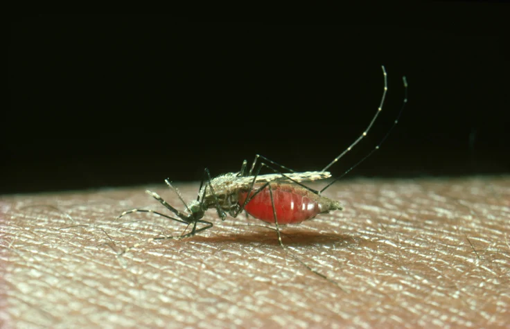
For nearly half a century, the malaria parasite Plasmodium falciparum has remained one of the most elusive and lethal adversaries in global health. Every 48 hours, inside the bodies of millions of people, a synchronized biological event occurs: trillions of parasites rupture from infected red blood cells and launch a frantic, coordinated assault to invade new ones. Central to this process is the "moving junction" (MJ)—a mysterious ring-shaped structure that scientists have known to be essential for invasion since the late 1970s.
Until now, the MJ remained a "black box." Because the structure assembles, functions, and dissipates within a fleeting 60 to 90 seconds, it has defied all attempts at detailed observation. However, a landmark study from Columbia University, published in the journal Cell, has finally captured this transient machinery in high-resolution, three-dimensional detail. The findings not only solve a decades-old biological mystery but also provide a structural blueprint for a new generation of life-saving antimalarial drugs.
The Mystery of the Moving Junction
The malaria parasite’s lifecycle is a masterpiece of evolutionary adaptation, involving two hosts—humans and Anopheles mosquitoes—and multiple physiological environments, including the liver and the bloodstream. The most critical point of vulnerability is the moment the parasite forces its way into a human red blood cell (RBC).
In 1978, electron microscopy first revealed a peculiar thickening at the contact point where the parasite meets the host cell membrane. Researchers eventually identified four key proteins—AMA1, RON2, RON4, and RON5—that form the building blocks of this junction. While it was clear that these proteins were essential for the parasite to breach the host’s defenses, their actual mechanics remained hidden.
"We’ve known for decades that this structure is essential for the parasite to get into a cell, but not how it actually works," explains Chi-Min Ho, PhD, an assistant professor in the Department of Microbiology and Immunology at the Columbia University Vagelos College of Physicians and Surgeons and the study’s senior author. "Efforts to address this critical gap in understanding have been thwarted by the short-lived nature of the complex, as well as by the difficulty of recapitulating it in heterologous systems."
Chronology of a Breakthrough: Freezing the Action
The Columbia team’s breakthrough relied on a bold strategy: stopping the parasite mid-stride. Recognizing that they could not study the MJ in a test tube, the researchers decided to "freeze" the invasion process in situ.
The team utilized a specific chemical compound designed to halt the parasite’s internal motor—the engine that drives its movement—without interfering with the assembly of the junction itself. By stalling the parasites precisely when they were halfway through the host membrane, the researchers were able to extract the intact AMA1-RON complex.
Once extracted, the complex was subjected to cryo-electron microscopy (cryo-EM). In this technique, samples are flash-frozen to preserve their native state and then bombarded with electron beams at extremely high magnifications. This allowed the team to visualize the structure in atomic detail for the first time.
The resulting images revealed a structure that defied existing scientific assumptions. Rather than a static, passive doorway, the MJ appeared to be a sophisticated molecular machine. Its shape, described by researchers as resembling a "sailboat," features the AMA1 protein acting as a "sail" above the cell surface, while the RON proteins form a broad "hull" pressed firmly against the host membrane.
Supporting Data: Mechanics of Invasion
The "hull" of this molecular sailboat held the most significant revelation. The surface of the RON complex is blanketed with positively charged anchors and studded with short, wedge-like helices.
"These short helices insert asymmetrically into one leaflet of the membrane, displacing lipid headgroups and applying lateral pressure to generate local membrane deformations," the authors noted in their report.

This discovery fundamentally shifts the understanding of how Plasmodium gains entry. For years, the scientific community operated under the hypothesis that the MJ was a series of "spot-welds" or a passive ring that the parasite simply pushed through. The data from the Columbia team demonstrates that the MJ is, in fact, an active, dynamic portal.
To confirm this function, the researchers performed a synthetic validation. They manufactured the wedge-like helices and introduced them to artificial membrane bubbles. The results were dramatic: the helices punctured and thinned the membranes, confirming that the structure is engineered to physically deform the host cell to facilitate entry.
"What we see instead is a machine built to reshape the host cell’s own membrane," says Meseret Haile, the study’s first author and a PhD candidate in the Ho lab. "That changes how we think about the whole event."
Implications for Global Health
The stakes for this research are exceptionally high. Malaria continues to claim the lives of approximately 600,000 people annually, with the vast majority of victims being young children in sub-Saharan Africa. Furthermore, the Plasmodium parasite is rapidly developing resistance to existing frontline therapies, rendering current antimalarial strategies increasingly ineffective.
Because the moving junction is conserved across different species and lifecycle stages of the parasite, it represents one of the most attractive targets in infectious disease research. If scientists can successfully block the MJ, they can halt the infection cycle at its most critical point of entry.
Using the high-resolution structural map generated by the cryo-EM analysis, the team employed machine learning-powered protein design tools to develop a "mini-protein." This synthetic agent was designed to break the grip between AMA1 and its partner protein, effectively disabling the "sailboat" machinery.
In laboratory trials, the mini-protein successfully blocked the invasion of red blood cells in a dose-dependent manner. Crucially, the treatment did not affect cells that were already infected, proving that the mechanism is specific to the act of entry rather than a general toxic effect.
Future Horizons: From Proof-of-Concept to Therapy
While the mini-protein is currently a "proof-of-concept" and not yet a clinical drug, the implications for future therapy are profound. The team’s methodology—capturing fragile, transient complexes directly from their native environment and using that data to guide the design of therapeutic binders—could serve as a blueprint for targeting other difficult-to-study pathogens.
"Once we could see the target in its real setting, designing something to block it became a tractable problem," says Daphne Kaxiras, an MD-PhD student in the Ho lab who spearheaded the inhibitor design. "That’s the part we’re most eager to build on."
The structural insights gained from this study also provide a clearer picture of how existing anti-malaria antibodies function. This information could be directly integrated into future vaccine development, allowing researchers to refine vaccines to better trigger the immune system to disrupt the MJ complex.
As the team looks toward the future, the goal is to refine these mini-proteins for potential clinical applications. The success of this project highlights a shift in modern pharmacology: moving away from trial-and-error chemical screening toward structural, precision-guided design. By finally pulling back the veil on the 60-second window of the moving junction, the Columbia researchers have opened a new door in the long, arduous fight against one of humanity’s oldest and most persistent killers.