Decoding the Hijacker: Scientists Map Influenza A’s Manipulation of Human Cells at Atomic Resolution
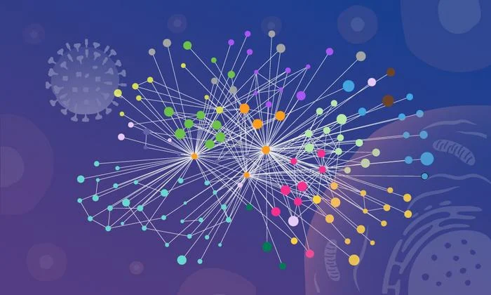
In a landmark achievement for molecular biology, researchers at EMBL Hamburg and the Leibniz Research Institute for Molecular Pharmacology (FMP) have achieved what was once considered a "holy grail" of virology: mapping the protein-level interactions between the influenza A virus (IAV) and its human host inside intact, living cells.
By combining the precision of in-cell cross-linking mass spectrometry (XL-MS) with the predictive power of AlphaFold structural modeling, the team has provided an unprecedented, high-resolution blueprint of how the flu virus systematically dismantles and repurposes human cellular machinery to fuel its own replication. This study, published in Nature Microbiology, not only sheds light on the stealthy strategies of the influenza virus but also establishes a scalable framework that could revolutionize the development of next-generation antiviral therapies.
The Silent Invader: A Global Health Burden
Every year, seasonal influenza exerts a staggering toll on global health. According to data from the World Health Organization and public health researchers, the virus is responsible for up to 650,000 deaths and millions of cases of severe illness annually. Despite the existence of vaccines and antivirals, the influenza A virus remains a moving target, constantly evolving to evade immune detection and develop resistance to standard treatments.
The core of the problem lies in the virus’s simplicity. Upon entering a human cell, IAV releases its viral RNA—a genetic blueprint for roughly 14 proteins. These viral proteins are not self-sufficient; they must "hijack" the host cell’s internal infrastructure to replicate. This process is entirely dependent on Protein-Protein Interactions (PPIs). By forging thousands of specific, often transient contacts with host proteins, the virus effectively converts a healthy cell into a virus-manufacturing factory.
Historically, scientists have struggled to visualize these interactions. Conventional biochemical methods required researchers to "lyse" or break open the cell, which destroyed the delicate, spatially confined compartments where these interactions occur. Without the architectural context of the cell, it was impossible to distinguish between meaningful viral-host interactions and "noise" created by proteins meeting only after the cell had been destroyed.
Breaking the Barrier: The Methodology of "Mapping in Context"
The breakthrough came through the development of a specialized workflow, pioneered by researchers at FMP Berlin, including Boris Bogdanow and Fan Liu. Their adaptation of XL-MS allowed the team to "freeze" protein interactions in place while the cell was still intact.
A Three-Pillar Approach:
- In-Cell Cross-linking (XL-MS): This technique acts like a molecular "glue." By adding cross-linking chemicals to living, infected cells, the researchers covalently bonded viral proteins to the host proteins they were touching at that exact moment. When the cells were later broken down for analysis, these bonds remained intact, preserving a record of the virus’s precise location and contact partners.
- Structural Modeling via AlphaFold: While XL-MS identifies which proteins are touching, it does not always reveal the precise physical geometry of the interface. The team integrated their cross-linking data directly into AlphaFold, an AI-driven protein structure prediction tool. By "feeding" the experimental data into the algorithm, they could predict the physical "docking" of viral and host proteins with remarkable accuracy.
- Functional Validation: The researchers confirmed their structural findings through targeted cell-biology assays, ensuring that the modeled interactions were not merely computational artifacts but were biologically essential for viral replication.
"This gives us insight into the interface between the virus and the human cell," explains Dr. Boris Bogdanow, now a junior research group leader at the Institute of Virology at Charité – Universitätsmedizin Berlin. "By capturing short-lived and location-specific interactions, we can finally identify actionable targets for future pharmaceutical interventions."
Chronology of the Investigation
The project, led by Dr. Jan Kosinski of EMBL Hamburg and the Centre for Structural Systems Biology (CSSB), was a multi-year effort that moved from the broad observation of infection to the granular detail of protein mechanics.
- Initial Mapping: The team began by infecting human cell lines with the influenza A virus and applying the XL-MS workflow at various time points post-infection to capture the temporal nature of the viral lifecycle.
- Computational Synthesis: Once the mass spectrometry data was obtained, the researchers moved into the structural modeling phase. They specifically focused on the viral surface protein, hemagglutinin (HA), and its journey through the host cell’s internal transport system.
- Discovery of the Paraspeckle Hijack: During the analysis, the team observed a consistent pattern: the systematic dissolution of "paraspeckles," which are membrane-less, droplet-like organelles in the cell nucleus.
- Validation across Strains: To ensure this was a fundamental mechanism and not an anomaly of a single laboratory strain, the researchers tested multiple influenza variants across different cell lines. In every instance, the dissolution of paraspeckles occurred, confirming it as a core viral strategy.
Key Findings: The Anatomy of Hijacking
The study highlighted two primary ways in which IAV commandeers the host, providing deep insight into previously mysterious viral mechanisms.
1. Manipulating the Protein Processing Factory
The researchers traced the trajectory of the viral protein hemagglutinin (HA), which the virus uses to bind to and enter host cells. HA requires complex folding and glycosylation (the addition of sugar molecules) to become functional. The study revealed that IAV recruits specific host factors within the endoplasmic reticulum-Golgi system—the cell’s manufacturing and shipping center—to ensure that HA is correctly processed. Understanding these specific host "chaperones" provides a map for drugs that could block the virus from finalizing its own surface proteins.
2. The Paraspeckle Dissolution
Perhaps the most surprising finding was the virus’s targeted destruction of paraspeckles. These organelles are typically involved in regulating gene expression and cellular stress responses. By dissolving these compartments, the virus achieves two goals:
- Resource Liberation: It releases RNA-binding proteins that were trapped inside the paraspeckles, which the virus then repurposes to aid in its own RNA replication.
- Immune Suppression: By breaking down the structures responsible for the cell’s "alarm system," the virus effectively silences the host cell’s antiviral gene response, preventing the cell from signaling for help or triggering apoptosis (cell death) to contain the infection.
"Watching these tiny organelles in the nucleus dissolve, consistently across every cell line and every flu strain we tested, told us this isn’t a side effect of infection—it might be a strategy," says Dr. Iuliia Kotova, the study’s first author.
Implications for Future Therapeutics
The methodology developed in this study is likely to have an impact far beyond the study of influenza. By demonstrating that it is possible to map the structural "handshakes" between viruses and hosts in a living environment, the researchers have provided a blueprint for studying a wide range of pathogens.
Designing Better Drugs
Current antiviral drugs often target the virus directly, which frequently leads to the rapid development of drug resistance as the virus mutates. By focusing on the host proteins—the "essential machinery" that the virus requires to survive—researchers may be able to develop "host-directed" therapies. Because these host proteins are essential to the human cell’s baseline function, the virus cannot easily mutate to bypass them without also damaging its own ability to replicate.
Preparing for Pandemics
Dr. Bogdanow noted that while this study focused on a lab-adapted influenza strain, the workflow is highly adaptable. The team is already looking toward applying these methods to viruses of higher pandemic potential, such as the H5N1 avian influenza. By understanding the specific interaction networks that allow these highly pathogenic strains to thrive, scientists can identify the "weak links" in the virus’s strategy, potentially enabling the rapid development of broad-spectrum therapeutics.
A New Era of Structural Virology
The collaboration between EMBL, FMP, and CSSB represents a shift toward "systems-level" structural biology. By integrating data from disparate fields—mass spectrometry, AI-driven structure prediction, and classical cell biology—the team has successfully bridged the gap between the molecular scale and the cellular scale.
"Our work provides a new way to study flu-host interactions in their native context and with structural insight," says Dr. Jan Kosinski. "The current results are a snapshot of a moment during infection, but they open the door to studying flu-host interactions across the entire infection cycle."
As the scientific community continues to grapple with the threats of respiratory viruses, studies like this offer a crucial advantage: the ability to see the enemy in its own environment. By exposing the virus’s reliance on the host’s architecture, researchers are moving closer to a future where we don’t just react to the flu, but systematically dismantle its ability to invade our cells in the first place.