The Integrity Crisis: Redefining Standards for Western Blotting in Modern Research


Western blotting remains a cornerstone of life sciences research, providing an essential bridge between protein identification and functional analysis. Despite the rise of high-throughput proteomic technologies like mass spectrometry, the Western blot holds its ground due to its accessibility, cost-effectiveness, and unmatched ability to visualize specific protein-antibody interactions. However, this ubiquity has brought the technique under a microscope—not just for its scientific utility, but for its role in a growing reproducibility crisis. As major journals tighten their submission requirements to combat image manipulation, the scientific community is being forced to reconsider how it documents, processes, and normalizes its data.
The Pillars of the Problem: Why Data Integrity Matters
In recent years, the scientific literature has been marred by a wave of paper retractions, many of which stem from questionable image integrity in Western blot figures. These irregularities range from accidental over-processing to deliberate, malicious manipulation. The repercussions are severe: a single compromised figure can invalidate years of research, damage institutional reputations, and erode public trust in scientific findings.
Journals such as the Journal of Biological Chemistry, Cell, and Nature have moved to the front lines of this battle, implementing rigorous image-screening protocols. They are no longer merely asking for clean figures; they are demanding transparency. Researchers are now expected to provide "audit-ready" documentation that traces a signal from the raw membrane capture to the final, published image. This shift represents a fundamental change in the culture of laboratory research, moving away from "black-box" data presentation toward a paradigm of total transparency.
Chronology of a Technique: From Traditional to Transparent
The history of Western blotting is one of incremental innovation. Since its inception, the method has relied on the assumption that researchers would maintain rigorous controls. However, as molecular biology transitioned into a data-heavy, competitive field, the pressure to produce "clean" images led to the normalization of practices—such as excessive contrast adjustment or the selective cropping of bands—that border on, or cross into, scientific misconduct.
In the early 2000s, the emergence of digital imaging software gave researchers unprecedented control over their images. While this improved clarity, it also made manipulation easier and less detectable. By the 2010s, organizations like the Office of Research Integrity (ORI) began to flag Western blot manipulation as a primary source of data fabrication. Today, we are in the "Era of Accountability," where the burden of proof rests firmly on the scientist to demonstrate that every pixel in a published figure accurately reflects the underlying biological reality.
Understanding Data Misrepresentation
Data misrepresentation often begins in the "gray zone" of image processing. It is vital to distinguish between enhancement and manipulation.
The Gray Zone: Processing vs. Distortion
For presentation purposes, non-destructive adjustments such as global brightness and contrast shifts are often necessary. In many cases, background subtraction is a valid scientific requirement to account for membrane noise. However, these adjustments become problematic when they are applied inconsistently. If a researcher alters the contrast of a target band but not the loading control, they have effectively skewed the data.
Deliberate manipulations—such as duplicating bands to fill missing data, rotating bands to align them, or "stitching" lanes from different gels without clear, labeled demarcation—are egregious violations of research ethics. These practices can create the appearance of a clean, statistically significant result where none exists.
The Documentation Mandate
Nikolas Chmiel, PhD, of Bio-Rad Laboratories, emphasizes that the most practical defense against accusations of malpractice is thorough, contemporaneous documentation. To meet the evolving standards of high-impact journals, a researcher’s archive must include:
- The Original, Unprocessed Gel Image: The "ground truth" of the experiment.
- The Processed Publication Image: Clearly labeled with all adjustments made.
- Software Trail: Full disclosure of the image detection and processing parameters.
- Consistency Logs: Evidence that identical processing parameters were applied across all comparative samples.
The Role of Advanced Analysis Software
To facilitate this level of rigor, researchers are increasingly turning to specialized image analysis packages. ImageJ, the gold-standard tool developed by the National Institutes of Health, remains the preferred choice for many because it preserves raw data integrity while allowing for the creation of standardized macros. These macros are critical; they enable a researcher to apply the exact same processing sequence to every image in a dataset, eliminating the possibility of human bias.
For beginners, Fiji—an image processing package that bundles ImageJ with useful plugins and comprehensive documentation—is highly recommended. Other tools, including GIMP, Python with scikit-image, and commercial solutions like Image Lab, offer more specialized workflows. Regardless of the tool, the golden rule remains the same: quantitation must be performed on raw images within the linear range of detection, and macros must be validated against known standards to ensure reproducibility.
Normalization: The Crux of Quantitative Accuracy
While image presentation is crucial, the scientific validity of a Western blot ultimately rests on its normalization strategy. Normalization is the process of correcting for non-biological variability, such as differences in sample loading or transfer efficiency.

The Housekeeping Protein (HKP) Dilemma
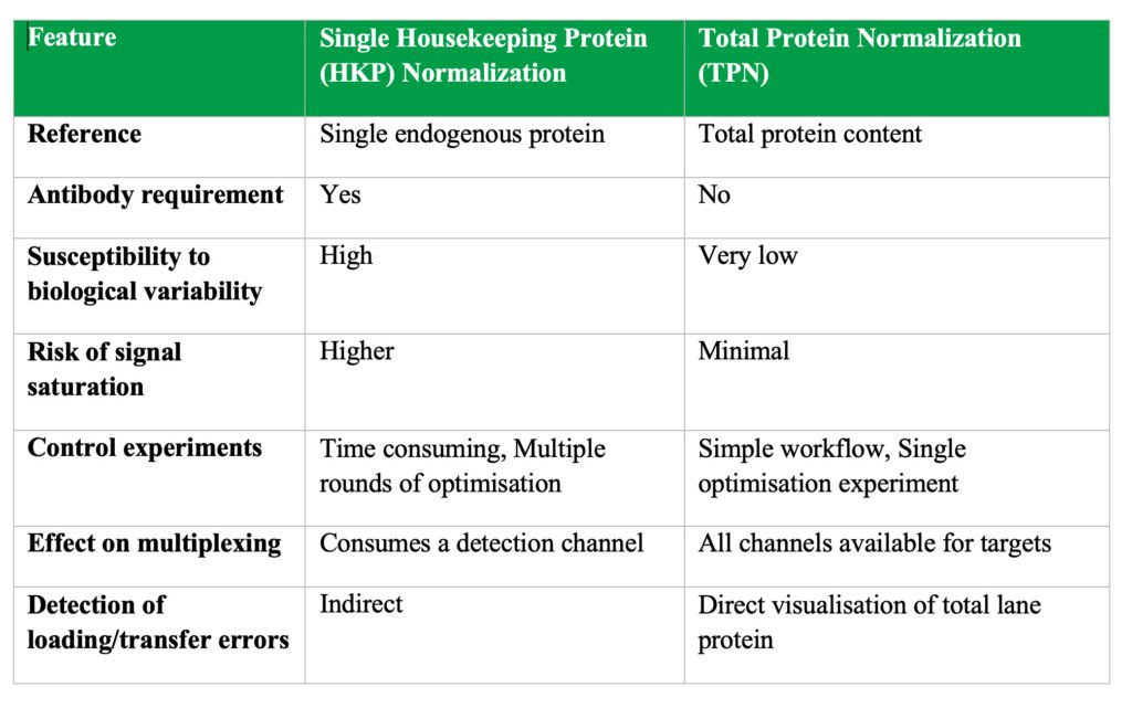
Traditionally, researchers have relied on "housekeeping" proteins like GAPDH, β-actin, or β-tubulin. The assumption is that these proteins are expressed at stable, consistent levels across all samples. However, this assumption is often flawed.
- Biological Variability: HKP expression can shift significantly due to cell state, drug treatment, or disease progression. In cancer research, for example, the expression of common HKPs can fluctuate wildly, rendering them unreliable as a standard.
- Linearity Challenges: HKPs are often highly abundant. If the signal is too strong, it can lead to oversaturation, where the detector cannot distinguish between varying protein amounts, causing the data to "plateau."
- Technical Error: The common practice of stripping and reprobing a membrane to detect both target and HKP can introduce significant error, potentially damaging the membrane and leading to incomplete signal removal.
The Case for Total Protein Normalization (TPN)
Total Protein Normalization (TPN) is increasingly viewed as the "gold standard" for quantitative Western blotting. By quantifying the total amount of protein loaded in each lane—rather than relying on a single, potentially unstable HKP—researchers can create a robust normalization factor.
Technologies such as Stain-Free imaging allow for the visualization of all proteins on a gel or blot, providing a true scalar relationship between the amount of protein loaded and the resulting signal. Studies comparing TPN to traditional HKP methods have consistently shown that TPN offers higher precision and reliability. By using TPN, researchers can often reduce the sample size required for statistical significance by more than 50%, as the method eliminates much of the "noise" introduced by suboptimal housekeeping controls.
Implications for the Future of Research
The transition toward more rigorous documentation and normalization is not merely an administrative burden; it is a necessary evolution of scientific practice. As journals continue to tighten their policies, the "business as usual" approach to Western blotting is becoming untenable.
The New Standard of Proof
The implications for the research community are threefold:
- Institutional Change: Research institutions must invest in training programs that teach students and postdocs not just how to perform a blot, but how to document it in an "audit-ready" fashion.
- Technological Adoption: Laboratories must move away from the "quick and dirty" HKP methods in favor of TPN and validated digital imaging software.
- Journal-Led Reform: Journals will likely continue to increase their scrutiny of raw data. Researchers should expect that submission without raw image files will eventually be rejected outright by most high-impact publications.
Conclusion
The Western blot is far from obsolete. Its capacity to provide meaningful, protein-level insights remains vital to our understanding of biology. However, the integrity of the data produced by this technique depends entirely on the scientist’s commitment to transparency. By adopting rigorous documentation practices, utilizing standardized analytical software, and transitioning toward more reliable normalization methods like TPN, the scientific community can restore the credibility of Western blot data. In an era where reproducibility is the ultimate measure of success, the ability to prove the authenticity of one’s results is just as important as the discovery itself.