Decoding the Survivor: How a New Mitochondrial Map Could Defeat a Formidable Pathogen
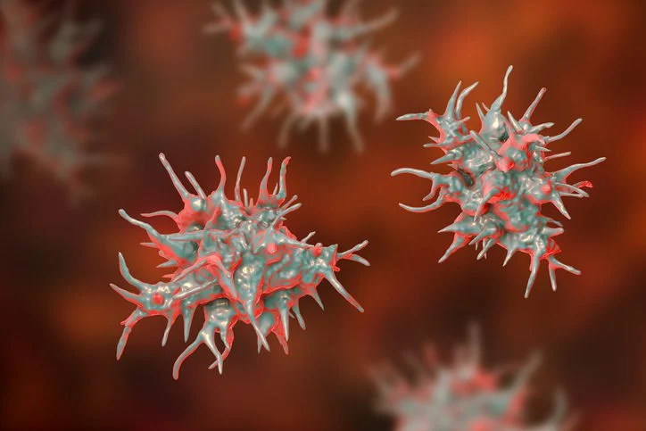
In the microscopic world, few organisms are as resilient—or as elusive—as Acanthamoeba. A ubiquitous, single-celled eukaryote found in the soil, air, and water, Acanthamoeba is generally harmless to the healthy human population. However, when it finds its way into the human eye, it can trigger Acanthamoeba keratitis, a devastating and sight-threatening infection.
For decades, clinicians have struggled to treat this condition effectively. The pathogen is notoriously difficult to eradicate, capable of transforming into a dormant, drug-resistant cyst when threatened by environmental stress or chemical assault. Current treatment protocols are not only limited in efficacy but are often toxic to human tissues. Now, a groundbreaking study published in the journal Cell—part of the ambitious MitoCarta Tree of Life Consortium—has unveiled a comprehensive map of the organism’s mitochondrial machinery. By identifying unique metabolic vulnerabilities, researchers are finally shedding light on how to neutralize this stubborn survivor.
The Chronology of a Scientific Breakthrough
The journey to mapping the Acanthamoeba "mitoproteome" was a multi-year undertaking that required a fundamental reassessment of the organism’s genetic blueprint.
Phase 1: Clarifying the Genetic Blueprint
The project began with a significant hurdle: the quality of the existing genome annotation for Acanthamoeba castellanii. Because the vast majority of mitochondrial proteins are encoded by the cell’s nuclear DNA, an accurate map of the mitochondria was impossible without first understanding the nucleus.
"If you imagine a page of words in a book, it would be like all the words were squished together and in a language that you don’t know," explained Dr. Jon Stefely, a metabolism investigator at the Morgridge Institute for Research and assistant professor at the University of Wisconsin School of Medicine and Public Health.
Using long-read RNA sequencing and advanced empirical methodologies, the team elevated the genome annotation accuracy from a meager 52% to an impressive 98%. This "deciphering" process was the necessary foundation for identifying which proteins were being sent to the mitochondria.
Phase 2: Building the AcMitoCarta
With a high-confidence genome in hand, the team, led by Dr. Stefely during his time in the laboratory of Dr. Vamsi Mootha at the Broad Institute, employed a multi-pronged proteomic approach. They utilized mitochondrial immunoprecipitation, density-gradient purification, high-resolution microscopy, and sophisticated mass spectrometry.
By observing which proteins became enriched as mitochondrial samples reached higher levels of purity, the researchers constructed the "AcMitoCarta"—a high-confidence inventory of 1,122 proteins. This catalog serves as the first definitive functional map of the Acanthamoeba mitochondrial system, providing a "who’s who" of the proteins responsible for the organism’s bioenergetic adaptability.
Supporting Data: Metabolic Flexibility Under the Microscope
The most striking revelation from the AcMitoCarta is the organism’s ability to perform a radical metabolic "retooling" when oxygen levels fluctuate.
The Hydrogen Gas Switch
Most organisms rely on standard aerobic respiration, where mitochondria consume oxygen to produce energy. However, the study confirmed that when Acanthamoeba is starved of oxygen, its mitochondria do not simply go dormant. Instead, they shift to a specialized anaerobic pathway. The researchers demonstrated that the pathogen activates a pyruvate:ferredoxin oxidoreductase-to-hydrogenase pathway, allowing it to produce hydrogen gas as a metabolic byproduct.
This oxygen-sensitive hydrogenase pathway is a key survival strategy. It allows the organism to persist in low-oxygen niches—such as the environment within an infected cornea—where other microbes might perish.
Uniqueness as a Therapeutic Target
The proteomic profiling revealed that of the 1,122 proteins identified, 381 have no recognizable counterparts in human or yeast mitochondria. More than 300 of these are entirely unique to the Acanthamoeba lineage.
This divergence is the "holy grail" of drug discovery. In pharmacology, the most effective drugs are those that target a biological process unique to the pathogen, leaving the host’s own cellular machinery untouched. Because these 300+ proteins do not exist in human cells, they represent a fertile field for the development of highly selective, non-toxic therapies.
Official Perspectives: The Clinical Challenge
The clinical reality of Acanthamoeba infections remains dire, as underscored by Dr. Stefely. Current treatments are often "blunt instruments"—chemical agents that lack specificity and can cause collateral damage to the delicate cells of the human cornea.
"It’s a big challenge. The lack of readily available diagnostics is a problem, and then even after you finally get to the diagnosis, we don’t have good drugs," Dr. Stefely noted. "Our current drugs are untargeted and toxic, and we just need more options for treating these infections."
The work published in Cell and its companion paper in Cell Press Blue—which focused on the organism’s encystation and the identification of cyst-wall proteins—represent a paradigm shift in how we approach this pathogen. Rather than guessing which drugs might work, researchers now have a "roadmap" of the proteins that are essential for the organism’s survival during its most dangerous phases.
Implications for Future Medicine
The implications of this research extend far beyond the laboratory. By mapping the mitochondrial proteome, the team has provided a framework for a new generation of targeted therapeutics.
Moving Toward Precision Therapy
The long-term goal for the Morgridge Institute and their collaborators is to systematically annotate the functions of every protein in the AcMitoCarta. By testing which of these proteins are essential to cyst formation or oxygen adaptation, researchers can identify "chokepoints" in the amoeba’s life cycle.
If a specific protein is required for the organism to form its protective, drug-resistant cyst, a drug that inhibits that protein could theoretically force the amoeba to remain in a vulnerable state, making it susceptible to the body’s natural immune defenses or existing antimicrobial treatments.
A Foundation for the Future
The MitoCarta Tree of Life Consortium, conceived by Dr. Vamsi Mootha, is demonstrating that by mapping the mitochondrial proteome across diverse species, we can unlock secrets of evolution and medicine simultaneously. The Acanthamoeba study is a proof-of-concept that this approach works even for the most difficult-to-treat pathogens.
As the scientific community begins to digest this new data, the path forward is clear:
- Targeted Inhibition: Developing small-molecule inhibitors that selectively bind to the 300+ unique mitochondrial proteins.
- Diagnostic Development: Utilizing the newly defined protein markers to create rapid, accurate diagnostic tests for clinical settings.
- Encystation Prevention: Specifically targeting the proteins that govern the transition into the dormant cyst state, preventing the pathogen from "hiding" from medication.
While the AcMitoCarta does not provide an immediate "cure-in-a-box," it fundamentally changes the terrain of the battle. For the patient suffering from a sight-threatening infection, the transition from "guesswork medicine" to "precision molecular targeting" is the most promising development in years. The resilience of Acanthamoeba has long been its greatest weapon; now, thanks to the high-resolution mapping of its own energy factory, that resilience may finally be its undoing.
With collaborators spanning the fields of structural biology, imaging, and mass spectrometry, the team is set to explore these vulnerabilities one set at a time. The era of the "unbeatable" amoeba is drawing to a close, replaced by a new era of molecular precision.