Breakthrough in Ocular Medicine: Duke Researchers Successfully Engineer Retinal Blood Vessels from Stem Cells
In a landmark achievement for regenerative medicine and ophthalmology, a team of biomedical engineers at Duke University has successfully generated specialized retinal endothelial cells (RECs) from human induced pluripotent stem cells (iPSCs). This development, published in the journal Nature Biomedical Engineering, offers a revolutionary pathway for both the treatment of debilitating retinal vascular diseases and the creation of highly accurate, human-based disease models.
The study, titled "Derivation of functional retinal endothelial cells from human pluripotent stem cells for therapeutics and modeling," represents the first time scientists have been able to reliably manufacture these critical cells, which form the delicate inner blood-retina barrier (iBRB) responsible for protecting the eye’s most energy-demanding tissue.
The Core Challenge: Understanding the Inner Blood-Retina Barrier
To appreciate the significance of this breakthrough, one must understand the biological complexity of the retina. Often described as the "window into the soul," the retina is, in biological terms, an extension of the central nervous system. Because it is tasked with the constant, high-energy process of converting light into neural signals, the retina requires a massive supply of oxygen and nutrients.
To support this demand, the eye maintains a specialized structure known as the inner blood-retina barrier (iBRB). This barrier is a sophisticated, highly selective biological sieve that regulates the passage of oxygen, water, ions, and nutrients, while simultaneously blocking harmful pathogens and toxins.
The structural integrity of this barrier depends on a specialized network of retinal endothelial cells (RECs), which work in tandem with pericytes and astrocytes to form tight junctions. When these junctions fail—a hallmark of diseases like diabetic retinopathy—the resulting vascular leakage and ischemia lead to progressive vision loss. Until now, researchers have been hampered by the inability to replicate these specific cells in a laboratory setting, as they do not exist anywhere else in the body and are notoriously difficult to isolate from human patients.
A Chronology of the Discovery
The journey toward this innovation began with the recognition that current methods for studying retinal vascular health were insufficient. Traditionally, researchers relied on cells derived from human patients, which are limited in supply, prohibitively expensive, and carry high levels of biological variability.
Phase 1: Reprogramming the Source
The research team, led by Sharon Gerecht, PhD, the Paul M. Gross Distinguished Professor and Chair of Biomedical Engineering at Duke, turned to iPSCs. These are mature adult cells that have been "reprogrammed" to return to a primal, pluripotent state, capable of differentiating into virtually any cell type in the human body.
Phase 2: The Biochemical Cocktail
Under the guidance of co-first authors Parker Esswein, a PhD student, and Dr. Ying-Yu Lin, the team established a rigorous protocol. They first coaxed the iPSCs into becoming generic endothelial cells—the building blocks of the vascular system. The critical innovation, however, was the subsequent application of a specialized "cocktail" of growth factors. By targeting the Wnt-β-catenin signaling pathway—specifically utilizing Norrin-Frizzled4 signaling—the researchers successfully guided the cells to adopt the unique identity of retinal endothelial cells (iRECs).

Phase 3: Benchtop Validation
With the iRECs successfully derived, the team moved to test their functionality. They placed these cells into 2D and 3D cultures to see if they would self-assemble into the complex, interconnected networks found in the human eye. The cells not only formed the necessary structures but also demonstrated the ability to form functional, tight-junctioned barriers.
Phase 4: Modeling Disease
To prove the clinical relevance of these iRECs, the team subjected them to "stress tests" designed to mimic diabetic retinopathy (DR). By exposing the lab-grown tissue to high glucose and low oxygen environments, the researchers observed the rapid breakdown of the barrier, mirroring the pathology seen in patients.
Phase 5: Therapeutic Application
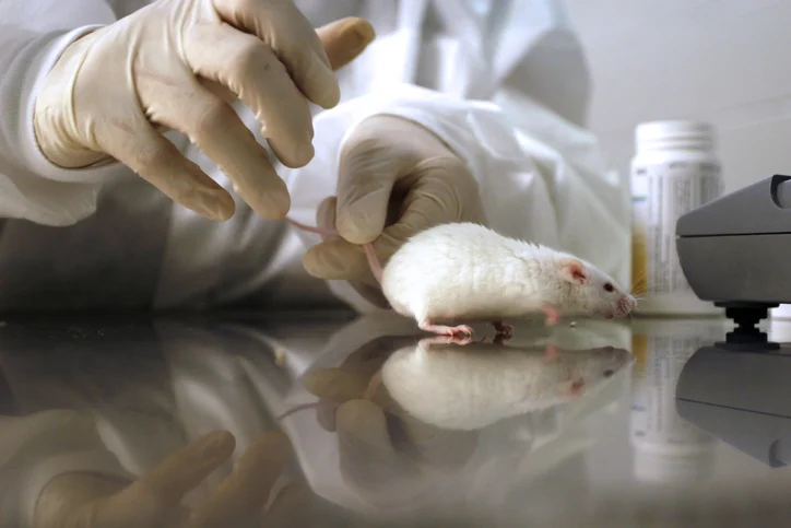
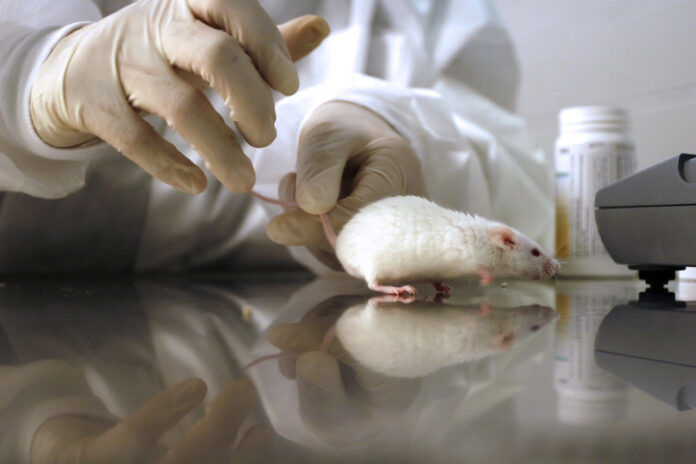
Finally, the team performed in vivo experiments using mouse models of retinal ischemia. By injecting the iRECs into the eyes of mice, they found that the cells successfully integrated into the existing vasculature, effectively "repairing" the network and revascularizing the ischemic tissue.
Supporting Data: Why This Changes the Landscape
The data generated by the Gerecht laboratory provides a roadmap for the future of ophthalmological research.
- Scalability: Unlike patient-derived cells, iPSCs provide a nearly infinite, consistent, and cost-effective supply of material. This allows for high-throughput drug screening, where researchers can test thousands of potential therapeutic compounds simultaneously.
- Precision Modeling: The ability to recreate the DR phenotype in a dish means that scientists can now observe the cellular mechanisms of eye disease in real-time. This is particularly vital for studying the early stages of diseases like diabetic retinopathy, which are often difficult to detect in living patients until permanent damage has occurred.
- Integration Potential: The successful revascularization in mouse models suggests that cell-based therapy could one day move beyond simple maintenance to active restoration of vision. By replacing dead or damaged endothelial cells with healthy, lab-grown versions, the researchers believe they can stabilize the retina and prevent the progression of blindness.
Official Perspectives: The Path Forward
Dr. Sharon Gerecht emphasized the urgency of the team’s work, noting that while millions of Americans suffer from retinal vascular diseases, our fundamental understanding of these conditions has remained stagnant for decades.
"Retinal vascular diseases affect millions of people in the U.S., but our understanding remains limited, hindering our ability to discover and develop new therapeutics," Dr. Gerecht stated. "Using human stem cells, we generated the cells found in retinal blood vessels, paving the way for new therapeutic approaches."
Parker Esswein, whose work on the project formed a core component of his doctoral research, highlighted the dual-purpose nature of the discovery. "When this specialized blood vessel tissue begins to break down, it can cause a lot of different diseases that lead to vision loss," Esswein remarked. "While there are sources of retinal endothelial cells, being able to grow a continuous supply from scratch could offer many advantages for those working in the field."
The team has already taken steps to ensure this technology can reach the commercial and clinical sectors, having filed a patent application covering both the iREC therapeutic method and the in vitro disease modeling platform.
Implications for the Future of Ophthalmology
The implications of the Duke University study extend far beyond the laboratory bench.
1. Accelerating Drug Discovery
The current model of drug development for eye diseases is slow and often relies on animal models that do not perfectly replicate human biology. With the availability of human-derived iREC models, pharmaceutical companies can perform more accurate preclinical trials, potentially reducing the time and cost required to bring life-changing drugs to market.
2. Personalized Medicine
Because iPSCs can be derived from a patient’s own skin or blood cells, this technology opens the door to "personalized" retinal models. Researchers could potentially derive iRECs from a specific patient with a rare genetic eye disorder to determine which drugs are most likely to be effective for that specific individual, minimizing the risk of adverse reactions.
3. Regenerative Therapies
While the current study focused on mouse models, the successful integration of iRECs into host vascular networks suggests a long-term future where clinicians might inject these cells into patients to treat conditions like diabetic retinopathy, macular degeneration, or retinal vein occlusion. By restoring the integrity of the inner blood-retina barrier, doctors could potentially "rebuild" the architecture of the eye.
4. Broadening the Research Scope
While the current report focuses on the successful derivation and initial disease modeling of iRECs, the researchers are already looking toward the future. The team plans to explore how these cells interact with other ocular cell types, such as neurons and microglia, to understand the holistic health of the retina.
In their concluding remarks, the authors stated, "Our study establishes functional human iRECs and microphysiological iBRB models that facilitate mechanistic studies aimed at identifying therapeutic targets and promoting the revascularization of injured retinas, thereby supporting treatment advancement."
As the scientific community digests these findings, it is clear that the Gerecht lab has provided a vital missing piece of the puzzle. By mastering the biology of the retinal blood vessel, they have brought the medical field one step closer to curing the leading causes of vision loss, offering a brighter outlook for millions of people worldwide.