Decoding the Architecture of Life: A Landmark Epigenetic Atlas Unveils the 3D Secrets of the Human Genome
In a milestone for genomic medicine, an international coalition of scientists led by the Salk Institute and the Arc Institute has unveiled the first body-wide, single-cell atlas mapping the dual landscape of the human epigenome. By simultaneously measuring three-dimensional (3D) genome folding and DNA methylation in the same cells, researchers have provided a comprehensive "instruction manual" for how human cells maintain their identity and function.
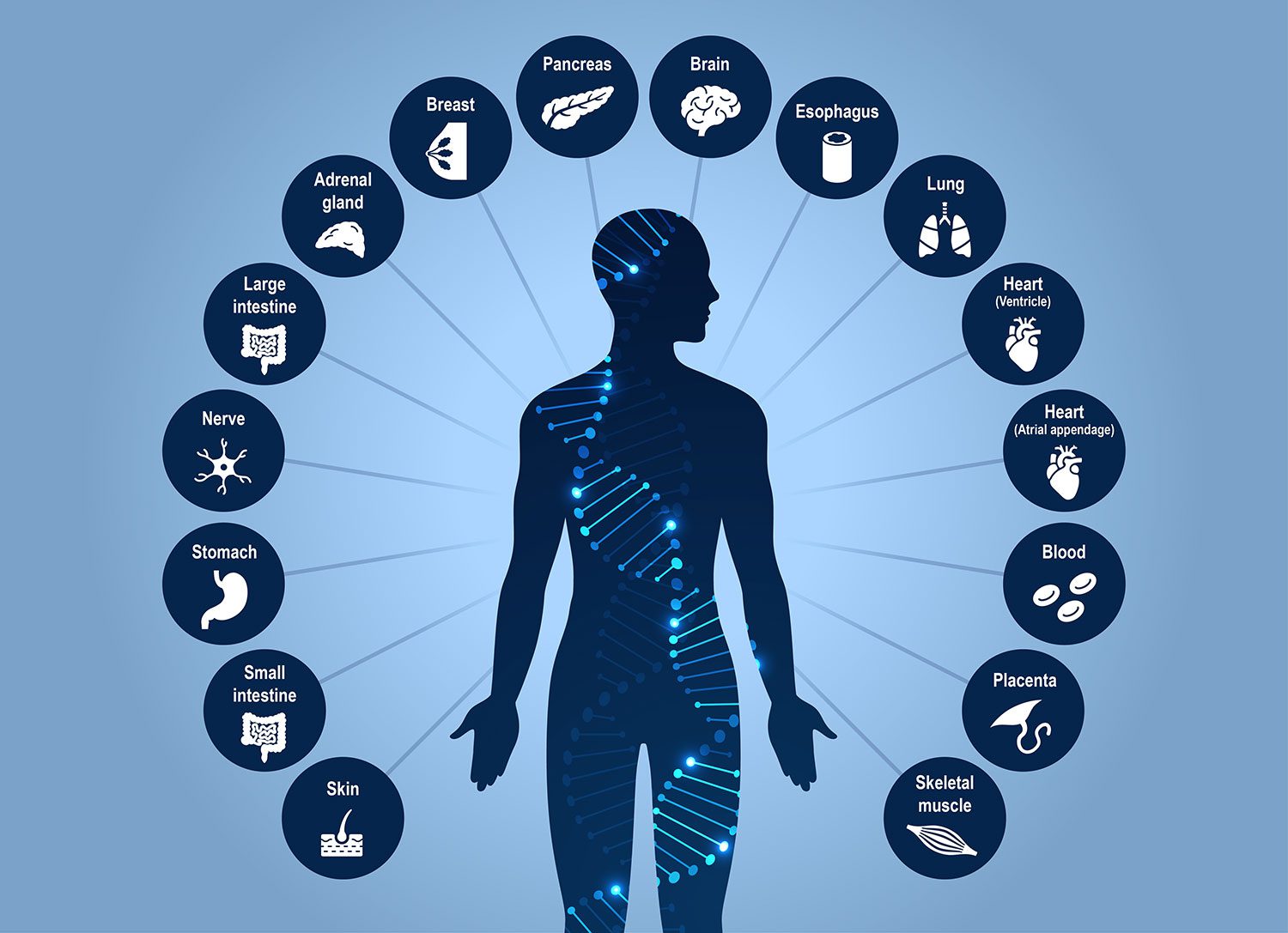
This expansive resource, which profiles 86,689 cells across 16 human tissues, was published as part of a major research package in Science and Science Advances. The findings offer a transformative perspective on how the linear string of DNA—long considered the primary code of life—is organized, folded, and chemically modified to create the vast diversity of human cell types.
The Main Facts: Beyond the Linear Code
The Human Genome Project, finalized in 2003, provided a foundational linear map of the three billion letters that comprise human DNA. However, this sequence alone fails to explain the biological complexity of the human body. A single genome must produce everything from the rhythmic cells of the heart to the complex excitatory neurons of the brain. The answer lies in the "epigenome"—the structural and chemical layer that governs how genes are switched "on" or "off."
The new study focuses on two critical components of this layer:
- DNA Methylation: A biochemical process where methyl groups are attached to DNA bases, effectively silencing specific genetic instructions.
- 3D Genome Organization: The physical architecture where DNA is looped and folded to bring distant genetic elements into contact, facilitating complex gene regulation.
For the first time, researchers were able to capture both of these features in the same individual cells, providing a high-resolution view of how cellular identity is physically and chemically encoded. The resulting atlas identifies 35 major cell types and 206 subtypes, providing an unprecedented level of detail that is now freely available to the global scientific community.
Chronology of Discovery: From Sequencing to 4D Mapping
The journey to this atlas is rooted in the National Institutes of Health’s (NIH) 4D Nucleome (4DN) program. The 4DN program was established with a specific, ambitious objective: to understand how the genome is organized in both space and time to regulate health and disease.
- Foundation Phase: Building on previous single-cell sequencing breakthroughs, researchers developed the multi-modal measurement techniques necessary to observe 3D architecture and methylation simultaneously.
- Execution Phase: Over several years, the team collected tissue samples ranging from the lungs and stomach to the heart and brain. These tissues were processed to extract 86,689 individual cells, which were subjected to rigorous epigenetic profiling.
- Analysis Phase: By overlaying these datasets with existing knowledge of genetic variants associated with diseases, the researchers began to bridge the gap between "junk" noncoding DNA and clinical pathology.
- Publication: In July 2026, the study, titled "Human body single-cell atlas of 3D genome organization and DNA methylation," was released alongside five companion papers in Science and three in Science Advances, marking a concerted global effort to standardize the understanding of the human 4D nucleome.
Supporting Data: Mapping the Noncoding Frontier
One of the most persistent hurdles in modern genetics has been the mystery of noncoding DNA. Most disease-associated genetic variants are located in regions of the genome that do not code for proteins. Consequently, identifying which cell types are affected by these variants—and which genes are subsequently disrupted—has been notoriously difficult.
The new atlas bridges this divide by identifying over 1.36 million differentially methylated regions and 283,606 differential chromatin loops. By mapping these features to known disease risk loci, the researchers uncovered clear, localized signatures:
- Metabolic Health: Variants linked to blood-glucose regulation were found to be concentrated in endocrine cells.
- Cardiac Health: Atrial fibrillation variants were specifically associated with the 3D structural signatures of heart muscle cells.
- Dermatology: Genetic variants linked to male-pattern baldness were localized to skin fibroblasts.
- Neurological Disorders: Variants associated with bipolar disorder and schizophrenia were found to reside in the regulatory landscapes of specific excitatory and inhibitory neurons.
Furthermore, the study debunked the long-held assumption that "non-CG methylation"—a specialized form of epigenetic marking—is unique to the brain and stem cells. The atlas demonstrates that this mark exists across a wide variety of human tissues, including muscle and pancreatic cells, suggesting it is a universal, albeit low-level, feature of cellular identity.
Official Responses and Expert Perspectives
The project’s scope has drawn praise from across the biomedical community for its potential to accelerate clinical research.
"There has been an appreciation for trying to understand, at the individual cell level, how the genome is organized, so that we can get a better idea of how genetic variants impact disease," said co-corresponding author Joseph Ecker, PhD, a professor at the Salk Institute and a Howard Hughes Medical Institute investigator. "Some cell types may be more vulnerable than others to genetic variants, because the genome is organized differently in different cell types—and whether a variant matters can depend on that organization."
Jesse Dixon, MD, PhD, associate professor at Salk and co-corresponding author, highlighted the diagnostic potential of the data. "A lot of the genetic variation that predisposes someone to disease is in noncoding parts of the genome. By adding in the 3D genome aspect, we can potentially bridge that gap—connecting noncoding variations with the genes they affect in specific cells and tissues."
The study also provides critical training data for the burgeoning field of artificial intelligence in medicine. As AI models become increasingly sophisticated at predicting the impact of genetic variants, they require high-fidelity, labeled datasets. This atlas serves as a gold standard for these models, effectively removing a major bottleneck that has previously hindered the accuracy of computational genomics.
Implications: The "Mismatch" Theory and Aging
Perhaps the most intriguing finding is the discovery of "epigenetic mismatches." In tissues like skeletal muscle, researchers observed cells that possessed the 3D genomic folding of mature, differentiated cells but retained the methylation patterns of stem cells.
This suggests that 3D architecture may update more rapidly than DNA methylation during cell development, indicating that cells are not always in a static state but are often in a state of flux. This revelation could fundamentally redefine how we categorize "cell types" and how we track the progression of diseases like cancer, where cell states are notoriously unstable.
The utility of the atlas was further demonstrated in a companion study led by Bing Ren, PhD, of the New York Genome Center and Columbia University. By utilizing the atlas’s cross-tissue methylation data, Ren’s team discovered that microglia—the brain’s primary immune cells—are replaced by cells resembling peripheral blood monocytes between the ages of 50 and 75.
"DNA methylation patterns are specific to each cell type and analogous to a cellular barcode," said Ren. "The comprehensive cross-tissue DNA methylation atlases show that the aging microglia in the human hippocampus more closely match the monocytes from peripheral blood than microglia from young adults, providing a crucial clue for the biological identity of these cells."
Future Directions: Moving Toward the Fourth Dimension
The current atlas provides a robust, static scaffolding for the human body, but as the name of the NIH program implies, the ultimate goal is to map these features across time. Future research will focus on the "fourth dimension": development, aging, and disease progression.
With 195 billion methylation measurements and 18 billion chromatin contacts now available through an interactive web browser, the resource empowers researchers to move beyond the linear constraints of the Human Genome Project. By providing a clear view of the structural and chemical layers that define a cell, the Salk and Arc Institute atlas offers a new roadmap for understanding the fundamental mechanics of human health and the precise origins of complex diseases. As science moves forward, this atlas will undoubtedly serve as the cornerstone for the next generation of precision medicine.