Decoding the "Zombie Cell": How MIT’s New RamanOmics Technology is Revolutionizing Aging Research
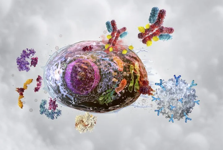
As the global population ages, the pursuit of "healthspan"—the number of years an individual lives in good health—has become a cornerstone of modern biomedical science. At the heart of this pursuit lies the mysterious world of cellular senescence. Often referred to as "zombie cells," senescent cells are those that have ceased to divide due to stress or DNA damage but refuse to undergo programmed cell death (apoptosis). Instead, they persist, lingering in tissues and secreting inflammatory molecules that degrade the surrounding environment, ultimately contributing to a host of age-related maladies ranging from osteoarthritis and type 2 diabetes to cancer and tissue degeneration.
A groundbreaking study published in Nature Aging has introduced a powerful new diagnostic framework known as "RamanOmics." Developed by researchers at the Massachusetts Institute of Technology (MIT), this technique promises to change how we detect, monitor, and potentially treat these elusive cells. By merging noninvasive optical imaging with sophisticated genetic profiling, the team has created a scalable, tissue-agnostic method to identify the "molecular architecture" of aging.
Main Facts: What is RamanOmics?
The fundamental challenge in senescence research has long been the trade-off between detail and viability. Traditional methods for identifying senescent cells—such as staining for p16 or p21 proteins—are inherently destructive; they require the fixation and death of the cells being studied. This makes it impossible to track the evolution of senescence over time in a living system or to observe these cells in their natural, undisturbed environment.
RamanOmics resolves this by combining Raman microscopy with single-cell gene expression data. Raman microscopy is a nondestructive optical technique that uses near-infrared or visible light to probe the biochemical composition of cells. When light interacts with a cell, the vibrations of its chemical bonds create a unique "spectral fingerprint."
By pairing this biochemical "optical biopsy" with transcriptomic data, the researchers created a high-resolution "barcode" for senescence. This barcode allows scientists to identify senescent cells based on their specific chemical signatures without the need for invasive sampling.
Chronology of the Discovery
The research, led by an interdisciplinary team from MIT, Harvard Medical School, and the Broad Institute, follows a multi-year effort within the National Institutes of Health (NIH) "Cellular Senescence Network" (SenNet).
- Initial Conceptualization: The team sought to bridge the gap between "what a cell is doing" (gene expression) and "what a cell is made of" (biochemical composition).
- The Pilot Study: Researchers utilized skin and lung tissue samples from two distinct cohorts of mice: young (2 months old) and aged (26 months old).
- Data Fusion: Using Raman microscopy, they captured the spatial vibrational signatures of thousands of cells. Simultaneously, they performed spatial RNA sequencing to map gene activity.
- Algorithm Development: By correlating the Raman spectra with the spatial RNA data, the team identified which biochemical bonds were most strongly associated with the genetic markers of senescence.
- Validation: The resulting "RamanOmics" barcodes were tested for accuracy, confirming that these spectral markers could successfully differentiate between healthy, young cells and senescent, aged cells.
- Publication: The findings were formally published in Nature Aging in September 2026, setting the stage for future clinical translation.
Supporting Data: Uncovering Tissue-Specific Aging
The study revealed that senescence is not a uniform process across the body. By comparing lung and skin tissue, the researchers uncovered fascinating, tissue-specific signatures of aging.
Metabolic Shifts and Lipid Accumulation
One of the most striking findings across both skin and lung tissue was a significant increase in lipid synthesis and accumulation in older cells. While the precise physiological impact of this "lipid loading" remains under investigation, it represents a clear biochemical hallmark of the aging process.
Skin vs. Lung: Unique Profiles
The researchers noted that senescence manifests differently depending on the organ’s function:
- Skin: The data showed a distinct decline in pathways associated with muscle contraction, extracellular matrix (ECM) remodeling, and collagen maintenance. This aligns with the physical reality of sagging skin and slower wound healing in the elderly.
- Lung: In contrast, aged lung tissue exhibited a surge in gene activity linked to immune activation and chronic inflammation. This shift is likely a contributing factor to the increased susceptibility of older adults to respiratory inflammation and diminished epithelial renewal.
"Together, these results reveal tissue-specific aging patterns," the authors noted in their study. "Immune activation and vascular remodeling with diminished epithelial renewal in lung, versus metabolic decline and impaired ion homeostasis with partial preservation of epithelial programs in skin."
Official Perspectives: The Experts Weigh In
The project was a collaborative effort involving some of the brightest minds in bioengineering and molecular biology.
Dr. Jeon Woong Kang, a research scientist at MIT and co-senior author, envisions a future where this technology moves from the lab bench to the clinic. "You can imagine that one day we may develop an endoscope that can look inside your body and identify cellular senescence," he noted. Such an innovation would allow doctors to monitor the "biological age" of specific organs in real-time.
Dr. Peter So, director of the MIT Laser Biomedical Research Center (LBCR) and co-senior author, emphasized that the goal is not to eliminate all senescence, but to understand it. "Senescence is not just a pathological condition," So explained. "It plays a role in so many normal physiological conditions, such as embryonic development and tissue regeneration. The idea behind the NIH Cellular Senescence Network is to take a very comprehensive approach to identify senescent cells because they are a double-edged sword."
Dr. Jian Shu, an assistant professor at MGH and Harvard Medical School, highlighted the necessity of their dual-view approach. "Our idea was to look at many different features to characterize senescence. That’s why we wanted to combine both single-cell gene expression and Raman microscopy, so that we can characterize the senescence from two complementary views."
Implications: The Future of Geroscience
The implications of RamanOmics for medical science are profound, particularly in the emerging field of senolytics—drugs designed to selectively eliminate senescent cells.
Precision Diagnostics
Currently, clinicians lack a way to noninvasively "see" where senescent cells are accumulating in a patient. RamanOmics could enable the creation of diagnostic tools that detect early markers of aging before physical symptoms appear. By focusing on a few specific Raman bands identified as the most "informative" in the study, researchers believe they can simplify the technology for faster, high-throughput clinical applications.
Longitudinal Tracking of Therapies
For researchers developing anti-aging drugs, RamanOmics offers a "gold standard" for monitoring efficacy. Instead of relying on surrogate markers, scientists could use Raman imaging to observe whether a senolytic treatment is actually clearing out senescent cells in a tissue sample over time.
Technological Scaling
The current system is in its infancy; it takes roughly 30 hours to analyze a one-square-millimeter tissue sample. However, the MIT team is already working on a higher-speed version of their imaging system. The goal is to scale this technology to analyze larger samples rapidly, moving toward a future where "longitudinal tracking of senescence in translational contexts—such as skin aging, fibrosis, or wound repair—becomes a clinical reality."
A Shift in Paradigm
By moving away from "destructive" testing, the RamanOmics framework aligns with the shift toward personalized, precision medicine. As the researchers stated in their report, "Coupling biochemical readouts with transcriptomic programs also opens opportunities for high-throughput screening of lipid-pathway modulators."
In conclusion, the MIT team has provided more than just a new tool; they have provided a new language for the body to describe its own aging process. As the technology matures, it will likely become an indispensable asset in the fight against the chronic, age-related diseases that currently define the final decades of human life. Through the lens of RamanOmics, the "zombie cells" that once haunted our biology may finally be brought into the light, providing clear targets for the next generation of life-extending therapies.