Geometry as the Architect: How Physical Constraints Shape the Blueprint of Life
In a landmark study published in Nature Materials, researchers at the European Molecular Biology Laboratory (EMBL) have unveiled a fundamental mechanism that dictates how embryos transform from simple cell clusters into complex, organized organisms. The paper, titled "Boundary geometry controls a topological defect transition that determines lumen nucleation in embryonic development," bridges the gap between theoretical physics and developmental biology, demonstrating that the physical shape of a tissue is not merely a consequence of growth, but a primary driver of biological architecture.
Main Facts: The Physics of Embryogenesis
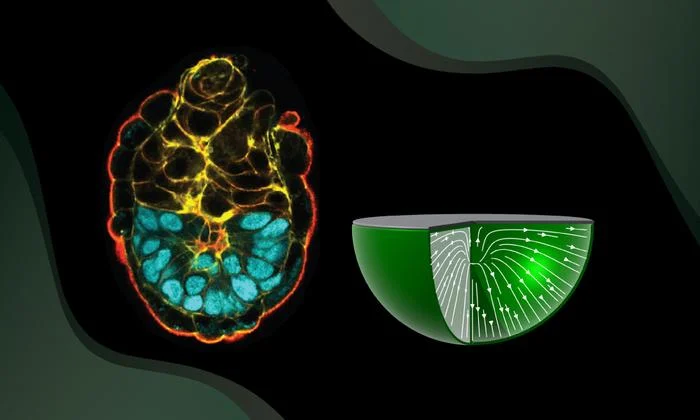
At the heart of early mammalian development lies the epiblast—a collection of polarized cells that serve as the "master architects" of the future body, destined to give rise to all major tissues. For years, biologists have understood that chemical signaling pathways govern cell differentiation. However, the EMBL team, led by group leader Anna Erzberger, shifted the focus toward the physical environment.
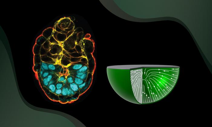
The study establishes that cells within the epiblast do not orient themselves randomly. Instead, their alignment is dictated by the boundaries of the tissue they inhabit. The research identifies a striking phenomenon: the presence or absence of an extracellular matrix (ECM) at the tissue boundary acts as a "switch" for cellular orientation. When cells interface with an ECM-lined boundary, they align perpendicularly; conversely, when they contact neighboring tissue directly, they align parallel to that border.
This interplay between cellular orientation and tissue geometry gives rise to "topological defects"—mathematical points of disruption where the orientation of the cells becomes undefined or singular. These defects are not errors; they are robust, predictable features of the system that serve as the nucleation sites for the formation of the lumen, a fluid-filled cavity essential for the structural integrity and subsequent development of the embryo.
Chronology: Bridging Disciplines
The path to this discovery was one of interdisciplinary synthesis. The project began with the observation that biological systems, while inherently "messy" and complex, often exhibit patterns that mimic the behavior of liquid crystals in physics.
- Theoretical Modeling (Phase 1): The team began by building a "minimal model"—a simplified mathematical framework designed to predict how polarized cells behave under varying physical constraints. By stripping away biological complexity, the physicists were able to isolate the role of geometry.
- Biological Validation (Phase 2): Working with mouse embryos, the researchers cross-referenced their mathematical predictions with live imaging. They mapped the orientation of cells within the cup-shaped epiblast, confirming that the model’s predictions matched the actual behavior of cells in situ.
- Perturbation Experiments (Phase 3): To prove causation, the researchers experimentally altered the geometry of the developing epiblast. By physically manipulating the embryo’s shape, they successfully induced the formation of additional lumina at locations predicted by their model, cementing the link between boundary shape and tissue formation.
Supporting Data: Understanding Topological Defects
The concept of a "topological defect" is central to this research. As co-first author Pamela Guruciaga, PhD, explains, these are points in space where the local order of the system breaks down.
"Imagine a starburst pattern of arrows," says Guruciaga. "The center of that pattern is a point where no single direction can be assigned. In the context of the epiblast, these points are incredibly robust. You cannot simply nudge them away; they are anchored by the geometry of the entire tissue."
The study provides quantitative evidence that the number and position of these defects are strictly controlled by the curvature and boundary conditions of the epiblast. By altering the "cup" shape of the tissue, the researchers demonstrated that they could effectively "program" where the lumen would form. This suggests that the embryo uses geometry as a high-level control parameter—a way to ensure that essential structures appear in the correct place every time, regardless of minor fluctuations in molecular signaling.
Official Responses: Insights from the Authors
The collaboration between physicists and biologists at EMBL highlights a growing trend in modern science: the use of "common languages" to decode life.
"For me, as a physicist, I may know why something works, but it’s still kind of magic to see that it’s all true in messy biological systems," remarked Dr. Guruciaga. "It was also super interesting coming from a pure physics perspective to come up with a common language to work with biologists."
Dr. Anna Erzberger, the group leader and co-corresponding author, emphasized the broader implications of the work. "What I find most exciting is that these results identify a very general physical principle," she noted. "We show that geometry alone can determine orientation patterns in three dimensions, independent of the microscopic details of the system. That means shape itself can act as a robust control parameter—not just in embryos, but across a wide range of biological and physical systems."
Implications: Beyond the Embryo
The implications of the EMBL study extend far beyond the early development of a mouse embryo. By proving that geometry acts as a fundamental "instruction manual" for cell organization, this research opens several new avenues for scientific inquiry:
1. Regenerative Medicine and Organoids
Current efforts to grow organoids—miniature, lab-grown organs—often struggle with achieving the correct structural maturity. If scientists can use the principles identified in this study to "mold" the geometry of growing tissues, they may be able to induce the formation of more functional, anatomically accurate organs for transplantation or drug testing.
2. Cancer Research
Tumors often involve a breakdown of the orderly cellular arrangements found in healthy tissue. Understanding the topological defects that govern healthy tissue architecture could provide new insights into how these structures fail, potentially leading to new diagnostic tools that identify the "geometrical signatures" of malignant growth.
3. Material Science
The principles discovered by the EMBL team are highly applicable to the development of "smart materials." Researchers designing synthetic tissues or robotic surfaces that can self-assemble could utilize these topological principles to create structures that change shape or function based on their environment.
4. Theoretical Biology
Perhaps most significantly, this study challenges the "gene-centric" view of development. While genes provide the blueprint for the individual proteins and cellular components, this research suggests that physics—specifically the physics of geometry and boundaries—provides the spatial constraints that allow those components to assemble into a coherent whole. It frames the embryo as a self-organizing system where geometry is not just a stage, but a lead actor.
Conclusion
The work of the EMBL researchers represents a paradigm shift in our understanding of life’s earliest stages. By demonstrating that the physical boundary of a tissue can dictate the behavior of cells through topological defects, the study provides a robust, elegant explanation for how order emerges from biological chaos.
As the scientific community continues to explore the intersection of physics and biology, studies like this serve as a reminder that the laws of the universe are as integral to our existence as our DNA. The "magic" of development, as Dr. Guruciaga put it, is grounded in the immutable laws of geometry—laws that dictate not only how an embryo forms, but potentially how all complex structures in the natural world take shape. Future research will undoubtedly focus on the limits of this principle: can we manipulate these defects to correct developmental anomalies? Can we scale this understanding to larger, more complex tissues? For now, the EMBL team has provided a definitive answer to a foundational question, proving that when it comes to the architecture of life, shape is substance.