A New Lens for Life: Laser Phase Plates Revolutionize Cryo-Electron Microscopy
For structural biologists, the challenge of visualizing the molecular machinery of life has long been akin to trying to read a newspaper through a thick, fogged-up window. While cryo-electron microscopy (cryo-EM) has revolutionized our ability to map the atomic architecture of proteins, the technique has been historically hampered by a fundamental physical barrier: the "fog" of low contrast, particularly when dealing with the smallest components of the human proteome.
Now, a breakthrough led by physicists at the University of California, Berkeley, and the Lawrence Berkeley National Laboratory has effectively wiped that window clean. By integrating a high-intensity "laser phase plate" (LPP) into a cryo-EM instrument, researchers have achieved a significant leap in image resolution, allowing scientists to visualize proteins that were previously too small or elusive to study. This development, recently published in the journal Science, promises to unlock the secrets of nearly 90% of the human proteome, potentially transforming our understanding of disease and drug discovery.
The Persistent Challenge: Why Small Proteins Remain Elusive
Since the 2017 Nobel Prize in Chemistry recognized the development of cryo-EM, the technique has become the gold standard for structural biology. It allows researchers to freeze samples in vitreous ice and image them with electrons, bypassing the arduous, and often impossible, task of creating high-quality crystals required for traditional X-ray crystallography.
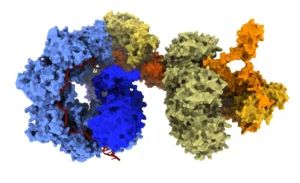
However, the physics of electron interaction creates a major bottleneck. Electrons, being charged particles, interact weakly with the atoms of small proteins. When a protein is below a certain size—roughly 70 kilodaltons (kDa)—the "signal" from the molecule is easily lost in the "noise" of the background. Because the vast majority of proteins involved in human biology fall below this size threshold, structural biologists have been effectively blind to a massive portion of the molecular landscape.
The struggle is not just about seeing a shape; it is about obtaining enough signal-to-noise ratio to discern the intricate folds and atomic details necessary to understand how a protein functions—or how it fails in the context of disease.
Chronology of an Innovation: From Concept to Reality
The journey to the laser phase plate began with a fundamental question in beam physics: how can we manipulate the electron wave without physical interference? Traditional phase plates—physical films placed in the beam path—often suffer from degradation, contamination, or unintended scattering, which can obscure the very data they are meant to reveal.

- Conceptualization (2018–2020): Dr. Holger Mueller and his team at UC Berkeley began exploring the use of light-matter interaction to shift the phase of the electron beam. They theorized that an intense, continuous-wave laser, if aligned with absolute precision, could act as a "virtual" phase plate.
- The Prototyping Phase (2021–2023): The team faced immense engineering hurdles. To achieve the necessary phase shift, the laser required a power density that would vaporize most materials. By utilizing highly polished, specialized mirrors to amplify the laser, they created a continuous-wave focal point of 75 kilowatts—a power level comparable to industrial welding lasers but condensed into a tiny, controlled focal point.
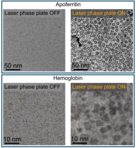
- Installation and Validation (2023–2024): The LPP was installed in a custom-configured Thermo Fisher Titan Krios microscope. Initial testing focused on "standard" test subjects like apoferritin and hemoglobin. The results were immediate: the LPP provided a dramatic boost in image clarity, particularly for the smaller, more challenging proteins that usually appear as indistinct blobs.
Technical Mastery: How the Laser Phase Plate Works
The LPP functions by fundamentally altering how the electron beam interacts with the specimen. In standard cryo-EM, the microscope relies on "defocusing"—purposely blurring the image—to generate contrast. While this makes the edges of a protein visible, it causes a catastrophic loss of high-resolution information.
The laser phase plate changes this paradigm. By creating a precise, high-intensity laser field, the system shifts the phase of the electron wave without requiring the microscope to be significantly out of focus. This "true" phase contrast allows the instrument to capture the high-resolution details of a protein’s structure while maintaining the visibility of its overall shape.
"The laser focus is 75 kilowatts focused to a few microns," Dr. Mueller explained. "That’s more power than what you use for welding. It has more power than a military laser. It builds up the brightest continuous laser focus ever."
By modulating the electron wave at the moment of passage, the system enhances the signal-to-noise ratio, effectively "sharpening" the image. This allows for superior specimen-motion correction—a process where researchers align thousands of individual images to reconstruct a 3D model—and more accurate classification of particle orientations.
Implications for Structural Biology and Cryo-ET
The implications of this technology extend far beyond single-particle analysis. One of the most anticipated applications is in cryo-electron tomography (cryo-ET).
While single-particle cryo-EM is used to study purified proteins in isolation, cryo-ET allows scientists to look at those proteins in their natural habitat: inside the crowded, chaotic environment of a living cell. In this context, the cell is a "forest," and the target protein is a single leaf. Finding that leaf has historically been an exercise in extreme frustration.
"With cryo-ET, we’re looking at small, very complicated cellular material that’s incredibly crowded inside the cell," says Bridget Carragher, PhD, founding technical director of imaging at the Chan Zuckerberg Biohub. "It’s like a forest of trees, and you’re trying to find one leaf on one tree in there. Cryo-ET needs a dramatic step forward in contrast, so we can start to see what’s going on inside the cell. That’s what the laser phase plate promises to give us."
By providing the necessary contrast to distinguish small proteins from the dense cellular milieu, the LPP opens the door to high-resolution "cellular atlases," where researchers can map the positions and interactions of proteins in real-time within the cell.
Official Perspectives: A "Step Function" Change
The scientific community has reacted to the publication with significant enthusiasm, recognizing the LPP as a potential paradigm shift in instrumentation.
Stephani Otte, PhD, vice president of imaging science at Biohub, believes the impact will be immediate. "This technology is a step function change for biology," she noted. "What was once invisible will become visible—and that changes everything about how we understand disease."
For pharmaceutical researchers, this means that the "druggable" genome—the set of proteins that can be targeted by small-molecule drugs—could expand significantly. Many of the most important proteins involved in cancer and neurodegenerative diseases are small, flexible, or found in low concentrations. If these proteins can be visualized at high resolution, the design of precision medicine will become significantly more efficient.
Dr. Mueller remains pragmatic but optimistic about the future of the technology. "The bottom line is, if you have a large protein and a really good sample, you may not need the phase plate to get a high-quality image. But for a small protein and a bad sample, laser-on is best."
Future Directions: Pushing the 17 kDa Threshold
The research team is not resting on their laurels. Having successfully demonstrated the technology with hemoglobin, they are now targeting even smaller molecules—aiming for proteins as small as 17 kilodaltons. Achieving this milestone would place almost every human protein within the reach of structural analysis.
Furthermore, development is already underway at the Biohub for a "dual-laser" system. This iteration is designed to further reduce component wear and minimize optical aberrations, ensuring that the system can be deployed more broadly in research environments.
As the technology matures and becomes more accessible to the wider structural biology community, the "fog" that has obscured the sub-70 kDa world will continue to lift. With this new, sharper lens, scientists are finally beginning to see the molecular foundation of life in unprecedented, crystal-clear detail. The era of the "invisible" protein is drawing to a close, and with it, a new, more precise era of biological discovery is beginning.