Rethinking the Building Blocks of Life: How Collagen’s Liquid State Rewrites Cell Biology
For over half a century, the scientific understanding of collagen—the most abundant protein in the human body—has been governed by a rigid, structural dogma. Textbooks have long depicted collagen as a long, stiff, rod-like molecule, essential for providing tensile strength to our skin, bones, tendons, and internal organs. However, a groundbreaking study from the Centre for Genomic Regulation (CRG) in Barcelona has shattered this paradigm, revealing that inside our cells, collagen exists not as a rigid fiber, but as a dynamic, liquid-like droplet.
This discovery, published in the Journal of Cell Biology, suggests that the fundamental mechanism by which our bodies produce and export their primary structural scaffolding is far more fluid and complex than previously imagined.
The Core Revelation: Collagen as a Liquid Condensate
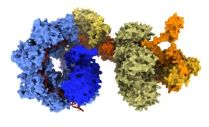
The research team, led by ICREA research professor Vivek Malhotra, utilized high-resolution live-cell imaging to observe the precursor form of type 1 collagen—procollagen 1 (PC1)—within the endoplasmic reticulum (ER). Type 1 collagen accounts for approximately 90% of the body’s total collagen, making its synthesis a critical operation for cellular health.
Under conventional microscopy, once collagen has left the cell and assembled into the extracellular matrix, it appears as rigid, extended rods reaching up to 400 nanometers in length. Because of this, scientists long assumed that collagen maintained this structure even while being synthesized inside the cramped confines of the ER.
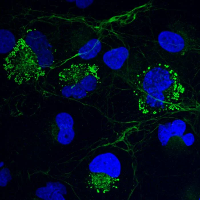
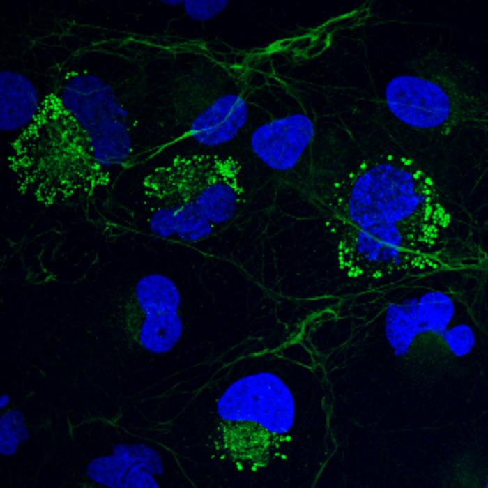
The CRG team’s findings challenge this directly. By observing human hepatic stellate cells—the liver cells responsible for collagen production and the primary drivers of liver fibrosis—the researchers discovered that PC1 gathers into small, spherical droplets. These droplets exhibit classic "phase-separation" behavior: they merge, split, and exchange material with their surroundings, much like oil droplets suspended in water.
"Inside a cell, collagens are not rigid molecules as one had assumed," said Dr. Vivek Malhotra, the study’s senior author. "They are, in fact, very pliable, taking a liquid condensate form much like oil in a drop of water."
A Chronology of Discovery: From Skepticism to Paradigm Shift
The path to this discovery was neither immediate nor accepted without intense scrutiny. The findings emerged from microscopy images captured by lead author Dr. Soumya Bhattacharyya in May 2024. Dr. Bhattacharyya was initially investigating the overproduction of collagen in fibrotic cells, a condition where excessive scarring occurs.
"I had no idea what it would lead to," Dr. Bhattacharyya recalled. "But when we took the samples, what struck me were these bright spherical structures you can’t miss."
When the team first shared these images within the laboratory, the immediate reaction was skepticism. The scientific dogma regarding protein folding and secretion is notoriously strict; cells have evolved elaborate quality-control mechanisms to detect and destroy misfolded or aggregated proteins, which can be toxic.
"I thought it must be an artifact," Dr. Malhotra admitted.
To validate the observation, the team spent months conducting rigorous follow-up tests. If these droplets were merely "junk" or misfolded proteins, the cell’s internal machinery—specifically a chaperone protein called BiP—would have been triggered to destroy them. Instead, the researchers found that the droplets were packed with helper proteins and chaperones that specifically recognize and facilitate the development of properly folded collagen. This confirmed that the liquid state was a functional, intended state of the protein, rather than a pathological error.
Supporting Data: Why "Rigid" Would Be Fatal
The implications of the liquid-like state go beyond mere aesthetics; it serves as a critical protective mechanism. If collagen were to assemble into its characteristic rigid, fibrous form while still inside the ER, the results would be catastrophic for the cell.
"This is another way by which cells ensure that collagens probably never become fibrous inside the cell," explained Dr. Malhotra. "Because if it were to become fibrous, it would kill the cell."
Furthermore, there is a logistical problem with the old "rigid rod" theory. The vesicles used to transport proteins out of the ER and toward the cell surface are typically only 60 to 90 nanometers in diameter. It has been a long-standing mystery in cell biology how a rigid 400-nanometer-long rod could be packaged and transported through such small cellular transit systems. By existing as a liquid condensate, collagen can be transported far more efficiently, bypassing the spatial limitations that a rigid structure would impose.
TANGO1 and the "Liquid Extrusion" Hypothesis
The study also provides critical insights into the role of TANGO1, a protein discovered by the Malhotra lab nearly two decades ago. It has long been known that TANGO1 is essential for collagen export, but its exact function remained elusive.
The new research clarifies that TANGO1 acts as a "mooring point." When the researchers depleted TANGO1, the collagen droplets still formed, proving that the protein is not required for the initial formation of the condensate. However, without TANGO1, the droplets were no longer anchored to the ER exit sites. Consequently, collagen secretion plummeted.
This suggests that the cell utilizes a "liquid extrusion" mechanism. Rather than relying on traditional, small transport vesicles, the researchers hypothesize that the collagen droplet attaches to the exit site and flows through it—a process similar to "wetting." Dr. Malhotra compares this to how plants move nutrients against gravity through capillary action, or how one might squeeze liquid out of a nozzle.
Broader Implications: Medicine and Disease
The paradigm shift in how we view collagen production has profound implications for medicine, particularly in diseases where collagen secretion is dysregulated.
1. Fibrosis
In conditions like liver, lung, and skin fibrosis, the body produces an excess of collagen, leading to the formation of dense scar tissue that impairs organ function. By understanding that collagen exists as a liquid condensate, researchers can now investigate ways to modulate this phase separation. If the process of "condensate formation" can be interrupted or slowed, it may offer a new therapeutic pathway to halt the progression of fibrotic diseases.
2. Oncology and Tumor Microenvironments
Perhaps the most striking implication involves cancer. Tumors often secrete massive amounts of collagen to create a dense, rigid extracellular matrix. This "tissue cement" acts as a physical shield, making it difficult for immune cells to infiltrate the tumor and preventing chemotherapy drugs from reaching their targets.
"One of the major problems in cancer is that the cells secrete so many collagens and other proteins out into the extracellular matrix that they hide in a shell," said Dr. Malhotra. "People are trying to find ways to break this tissue cement, and our study could help inform those strategies."
If scientists can develop drugs that either dissolve these liquid collagen droplets inside the cell or prevent them from mooring to the export sites via TANGO1, they could effectively starve the tumor of its protective shell, rendering it vulnerable to both the immune system and conventional medicine.
Future Directions
The team at the CRG is not stopping here. While the "liquid extrusion" hypothesis is currently a working model, the next phase of research will involve direct visualization of the export mechanism to confirm exactly how these droplets pass through the ER membrane.
Furthermore, the researchers are collaborating with external partners to develop a mouse model. This will allow them to observe these collagen droplets in living tissue, moving the study from the controlled environment of cell cultures to the complex, systemic reality of an animal model.
This research marks a significant departure from the established understanding of cellular secretion. By looking past the rigid fibers of the extracellular space and into the fluid heart of the endoplasmic reticulum, the team at the Centre for Genomic Regulation has opened a new chapter in cell biology—one that views the body’s most common protein not as a static rod, but as a fluid, dynamic, and highly regulated substance. As this model is tested and refined, it promises to reshape our approach to some of the most stubborn diseases in modern medicine.