Rewiring the Diseased Brain: New Breakthrough in Huntington’s Disease Research
Huntington’s disease (HD), a hereditary, neurodegenerative disorder, has long been viewed as a monolithic march toward cognitive and motor decline. While the genetic culprit—a trinucleotide repeat mutation in the Huntingtin (HTT) gene—has been understood for decades, the specific mechanics of how this mutation orchestrates the collapse of neural networks remained largely shrouded in mystery.
A groundbreaking study published in the journal Nature, titled "Restoring cortical disinhibition improves Huntington’s disease phenotypes," has fundamentally shifted this perspective. By identifying specific inhibitory neuron populations that malfunction during disease progression and using optogenetics to "re-tune" these circuits, researchers at UC San Diego and the Max Planck Institute for Biological Intelligence have demonstrated that the diseased brain may possess a latent capacity for recovery.
The Architecture of the Discovery: Main Facts
The research centers on the motor cortex, a region of the brain critical for orchestrating movement. In the past, neuroscientists largely focused on the death of excitatory neurons, often overlooking the role of inhibitory neurons. The prevailing assumption was that these inhibitory cells were spared from the ravages of Huntington’s.
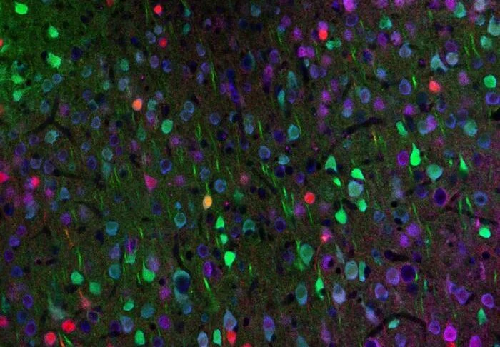
This study challenges that dogma. Researchers utilized advanced imaging techniques to track the real-time activity of individual cortical neurons in transgenic mice models of Huntington’s disease. They discovered that while the genetic mutation is systemic, the functional breakdown is highly specific: a profound imbalance of activity across different cell types.
Most notably, the team identified that vasoactive intestinal peptide (VIP) neurons—a specific class of inhibitory interneurons—showed a marked decline in activity as the disease progressed. These VIP neurons act as critical gatekeepers for neural plasticity; when they fail to fire, the brain loses its ability to adapt and refine its circuits in response to new experiences. By artificially stimulating these dormant VIP neurons, the research team successfully restored normal circuit activity and, crucially, improved the mice’s ability to learn and perform complex motor tasks.
A Chronological Progression of the Study
The road to this discovery was characterized by a meticulous transition from broad mapping to precise intervention.
Phase I: Mapping the Silence
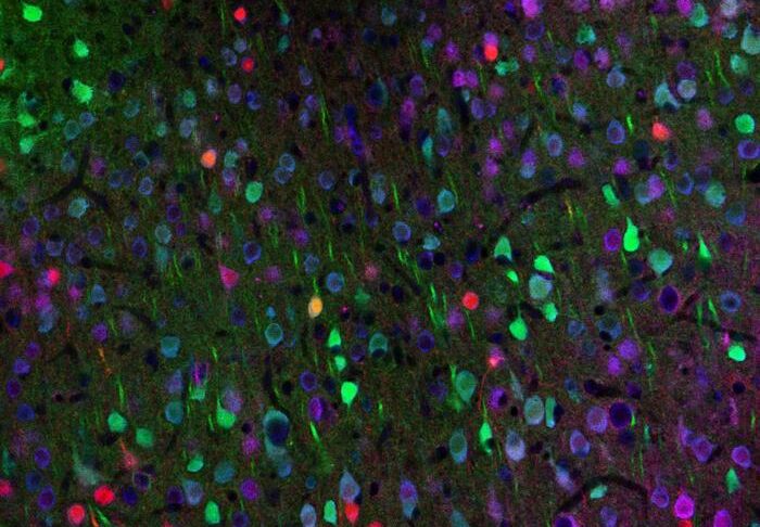
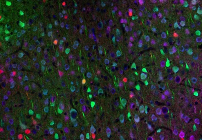
The research began by observing the longitudinal progression of HD in transgenic mouse models. Using high-resolution fluorescent labeling, the team monitored the motor cortex to see which neurons were firing normally and which were falling silent. This phase established the baseline "dysfunction profile" of the HD brain.
Phase II: Identifying the Vulnerable Gatekeepers
Upon identifying the underactive VIP neurons, the researchers needed to confirm their role. They observed that the silencing of these cells coincided with the onset of motor deficits. This shifted the hypothesis from "cell death" to "circuit imbalance." The researchers realized that the cells weren’t necessarily dying; they were simply failing to perform their inhibitory duty, causing a "noise" that prevented the motor cortex from learning new motor patterns.
Phase III: The Optogenetic Intervention
With the target identified, the team employed optogenetics—a biological technique that uses light to control the activity of specific cells that have been genetically modified to be light-sensitive. By shining pulses of light on the VIP neurons, the researchers were able to "kickstart" them back into a functional state.
Phase IV: Assessing Lasting Plasticity
Perhaps the most significant finding occurred after the stimulation sessions concluded. The researchers noted that the improvements in motor learning persisted for days after the light stimulation was turned off. This suggested that the intervention had not just masked the symptoms, but had triggered a lasting, regenerative change in the brain’s wiring.
Supporting Data and Technical Nuance
The study’s data offers a new lens through which to view neurodegeneration. In a healthy brain, VIP neurons perform a process called "disinhibition." By inhibiting the inhibitors, they effectively "open a gate" for excitatory neurons to facilitate learning.
In the Huntington’s-affected mice, this gate remained closed. The study quantified this by comparing the activity traces of VIP neurons against excitatory neurons during motor learning tasks. The data revealed that in the diseased state, the coordination between these two types of neurons was completely desynchronized.
The recovery metrics were striking:
- Correction of Activity Patterns: Stimulation of VIP neurons returned the overall cortical activity to levels statistically similar to those of healthy, wild-type mice.
- Behavioral Performance: The mice demonstrated a measurable increase in their ability to master motor tasks that were previously impossible for them to learn.
- Persistence: The "re-tuning" effect was not fleeting. Because the stimulation induced plasticity, the brain’s internal architecture remained optimized long after the optogenetic stimulus was removed.
Official Responses and Expert Perspectives
The study has sent ripples through the neuroscience community, offering a rare glimmer of hope for a condition that has historically lacked effective treatments.
"This work shows that correcting specific imbalances in brain circuits can restore function, even in a complex neurodegenerative condition," said Takaki Komiyama, PhD, a professor in the UC San Diego Departments of Neurobiology and Neurosciences. "It highlights the potential of targeting defined cell types to promote recovery rather than just trying to stop the initial genetic damage."
Dr. Irina Dudanova, a co-author of the study, emphasized the shift in how we must view cell loss in neurodegeneration. "Cortical inhibitory cells have received little attention in Huntington’s disease, as for a long time they were considered to be spared from neurodegeneration," she explained. "Surprisingly, we detected profound changes in their activity. If we know which cells to target, we can retune the brain’s abnormal activity patterns. This gives hope for future therapies."
Sonja Blumenstock, PhD, who led the behavioral analysis, added, "By activating the VIP inhibitory cell type, we gradually restored more normal activity patterns, and, very importantly, we also saw an improvement in the ability of the mouse to learn a motor task."
Implications for Future Medicine
The implications of this research extend far beyond Huntington’s disease. By proving that a "diseased" brain can be nudged back into a functional state through targeted circuit modulation, the researchers have opened a new frontier for treating various neurodegenerative and neurodevelopmental conditions.
Moving Toward Non-Invasive Therapy
While optogenetics currently requires genetic modification and physical light delivery, the researchers are looking toward the future. Dr. Komiyama envisions a time when non-invasive techniques—perhaps using ultrasound or electromagnetic fields—could achieve similar "tuning" of specific cell populations without the need for invasive brain surgery.
Redefining Neurodegeneration
The study provides a compelling argument for moving away from the "neuron death" model of disease. If researchers can identify the specific, underperforming nodes in a network, they may be able to restore function even while the underlying genetic defect persists. This "circuit-restoration" approach is likely to become a foundational concept in the next decade of neurology.
A Beacon for Learning and Recovery
Perhaps the most optimistic outcome is the discovery that the HD brain remains capable of learning. By "opening the gate" to plasticity, the team has suggested that the cognitive decline in Huntington’s may be partially reversible. As the team moves forward, they hope to translate these findings into clinical applications that could help human patients regain motor control and cognitive function.
"We have come up with a way to allow the diseased brain to learn better," concluded Komiyama. "Our hope is that a related approach will help people with impairment in their learning abilities, providing a pathway to recovery that was previously thought to be impossible."
This research marks a turning point in how we understand the brain’s resilience. Even as the shadow of a genetic mutation looms, the delicate, intricate dance of our neural circuits offers a potential pathway to reclaim what the disease attempts to steal. The work of the Komiyama and Dudanova teams serves as a testament to the idea that, in the brain, the circuits are not just hardware—they are a dynamic, living system that, with the right intervention, can be brought back into harmony.