Unlocking the Cellular Secrets of Diabetic Kidney Disease: New Insights from Organoid Models
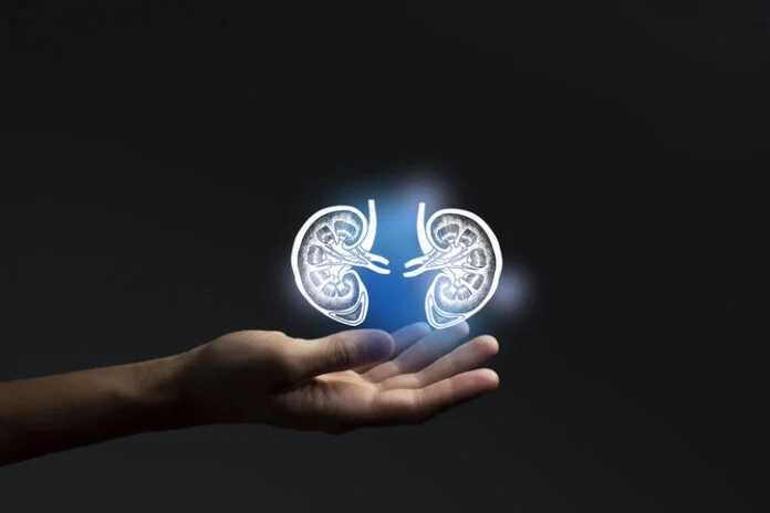
Diabetic kidney disease (DKD), a devastating complication affecting approximately 40% of individuals living with diabetes, has long been viewed through the lens of systemic circulatory stress. For decades, the medical consensus centered on the idea that chronic high blood pressure and systemic inflammation—driven by external immune cells—were the primary culprits behind the gradual destruction of the kidney’s filtration apparatus. However, a groundbreaking study published in Stem Cell Reports suggests a paradigm shift: the kidneys themselves may be capable of triggering their own inflammatory demise when exposed to sustained hyperglycemia.
By leveraging advanced human kidney organoids, researchers have discovered that elevated glucose levels can induce a "tissue-intrinsic" inflammatory response, leading to the catastrophic detachment of podocytes—the delicate cells essential for maintaining the kidney’s filtration barrier. This finding not only challenges existing models of DKD but also opens the door to novel therapeutic interventions that target the kidney’s internal molecular machinery rather than just systemic blood sugar levels.
Main Facts: The Hyperglycemic Trigger
The study, led by Benjamin S. Freedman, PhD, of the University of Washington and the University of Miami, provides a sophisticated look at the molecular mechanisms of DKD. The research team successfully demonstrated that human kidney organoids, when cultured in high-glucose environments, undergo significant morphological deterioration.
Crucially, the damage observed was not the result of direct cellular toxicity or cell death caused by glucose poisoning. Instead, the cells began to detach from the organoid body, mirroring the physical signs of kidney decline seen in human patients. Perhaps most surprisingly, this inflammatory response occurred in a "sterile" environment—the organoids lacked a vascular system and circulating immune cells. This proves that the kidney tissue itself is capable of mounting an inflammatory defense that, paradoxically, contributes to its own destruction.
Chronology of the Discovery
The research trajectory began with the need to bridge the gap between clinical observation and cellular pathology.
- Model Development: The team utilized pluripotent stem cells to engineer human kidney organoids through a stepwise differentiation process that mimics natural embryonic development. This provided a living, 3D architecture capable of responding to environmental stimuli.
- Controlled Exposure: Researchers subjected these organoids to varying concentrations of glucose to simulate the conditions of a diabetic patient.
- Observation of Deterioration: While organoids in low-glucose media remained healthy, those in high-glucose media exhibited progressive structural breakdown.
- Molecular Mapping: Using single-cell RNA sequencing (scRNA-seq), the team identified the specific gene expression signatures triggered by the sugar-rich environment.
- Validation via Inhibition: Finally, the team applied inhibitors for specific inflammatory pathways (MIF and TNF-alpha) to determine if they could reverse or halt the observed damage, confirming the causal role of these pathways in the disease phenotype.
Supporting Data: Decoding the Transcriptome
The power of the study lies in its transcriptomic analysis. By comparing the high-glucose organoids with data from human kidney biopsies, the researchers identified a striking overlap in gene expression.
The Role of MIF and TNF-alpha
Single-cell RNA sequencing pinpointed the Macrophage Migration Inhibitory Factor (MIF) as the most significantly upregulated gene across the dataset, specifically within the epithelial and podocyte clusters. Additionally, the study highlighted the activation of the TNF-alpha/NF-kB signaling pathway. In clinical practice, these markers are often associated with systemic immune responses; however, their presence in the organoids confirms that epithelial cells and podocytes are autonomously producing these inflammatory signals in response to hyperglycemia.
Assays of Cellular Integrity
To ensure the damage was not simply a result of cells dying from glucose exposure (cytotoxicity), the team employed lactate dehydrogenase (LDH) assays and live/dead staining. These tests confirmed that the cells were viable but had lost their structural adhesion—a phenomenon known as "detachment." This mirrors the clinical reality of podocyturia, where podocytes are shed into the urine of diabetic patients, signaling the breakdown of the glomerular filtration barrier.
Official Responses and Expert Perspective
Dr. Benjamin S. Freedman, the study’s senior author, emphasized the unexpected nature of the findings during a recent briefing. "High sugar causes inflammation in these organoids even though they lack an immune system," Freedman noted. "This was unexpected and gives us a new way to think about how diabetes can affect kidneys and other organs."
The research community has received the findings with significant interest. While the study is in its preclinical stages, experts in nephrology have pointed out that the ability to model "tissue-intrinsic" inflammation provides a level of granular control previously unavailable in animal models. By isolating the kidney tissue from the noise of systemic circulation, the researchers have created a "clean room" for testing drugs that could stabilize the filtration barrier before permanent damage occurs.
Implications: A New Frontier in Therapeutics
The discovery that the kidney’s own inflammatory pathways are active participants in DKD provides a roadmap for future drug development.
Targeting the "Inside-Out" Damage
Currently, standard care for diabetic patients focuses on systemic management—using ACE inhibitors to control blood pressure and SGLT2 inhibitors to manage glucose and renal stress. However, these treatments do not directly "stop" the molecular signaling that causes podocyte detachment. The current study suggests that targeting specific pathways—specifically MIF and TNF-alpha—could act as a protective layer, shielding the kidney from the inflammatory consequences of hyperglycemia.
Potential Therapeutic Agents
The study successfully demonstrated that:
- ISO-1 (a MIF inhibitor) protected the organoids from podocyte injury.
- Etanercept (a TNF-alpha inhibitor) similarly mitigated the damage.
- MAPK pathway inhibitors provided partial rescue of podocyte morphology.
These findings suggest that a multi-pronged pharmacological approach might one day be viable, where standard glucose control is augmented by "renal-protective" agents that specifically silence the kidney’s inflammatory alarm system.
Limitations and Future Directions
Despite the promising results, the research team is careful to temper expectations. The study highlights several key limitations that must be addressed before clinical trials can be considered:
- Hyperglycemic Thresholds: The model required higher-than-normal glucose concentrations to induce the injury phenotype, suggesting that the organoids might require additional triggers (such as mechanical stress or hormonal signals) to fully replicate the human disease state in vivo.
- Lack of Vasculature: The kidney is a highly vascularized organ. Without blood flow, the organoids cannot account for the hemodynamic pressures that contribute to DKD. Future iterations of this model will need to incorporate endothelial cells and perfusion systems to simulate the physical forces of the human circulatory system.
- Immune System Absence: While the study proved that inflammation can be intrinsic, the researchers acknowledge that in a living patient, this intrinsic inflammation likely "talks" to the systemic immune system. Future studies will need to introduce immune cells into the organoid environment to understand the cross-talk between the kidney and the body’s defense mechanisms.
Conclusion
The study published in Stem Cell Reports serves as a vital reminder that the cellular mechanisms of complex diseases often remain hidden in plain sight. By stripping away the complexity of the systemic immune response, Dr. Freedman and his team have revealed a fundamental vulnerability in the kidney: its own metabolic and inflammatory sensitivity to sugar.
As the scientific community moves forward, the use of organoids as "disease-in-a-dish" models will likely accelerate the discovery of targeted therapies. While a cure for diabetic kidney disease remains a distant goal, the shift toward understanding the kidney as an active, self-damaging participant in its own decline is a critical step. The potential to "silence" the kidney’s inflammatory response could eventually transition DKD from an inevitable progression toward failure into a manageable, and perhaps even preventable, condition.
For the millions of patients currently navigating the challenges of diabetes, these insights provide a glimmer of hope: that the next generation of kidney medicine will be defined not just by how we manage our blood, but by how we protect our tissues from the inside out.