Rewiring Vision: How Biomimetic Nanoparticles Are Offering New Hope for Retinal Degeneration
In the complex landscape of ocular medicine, the loss of photoreceptor cells—the specialized neurons in the retina that convert light into electrical signals—has long been considered an irreversible catastrophe. Whether caused by retinitis pigmentosa, age-related macular degeneration, or other degenerative conditions, the "death" of these light-sensing cells effectively cuts the communication line between the outside world and the brain.
However, a groundbreaking study published in Nature Biomedical Engineering suggests that the hardware of the eye may remain functional even when the "software" (the photoreceptors) has failed. An international team of researchers, led by Associate Professor Menglin Chen of Aarhus University, has successfully demonstrated that injectable, light-sensitive nanoparticles can act as a wireless bridge, restoring light sensitivity to degenerated retinas in preclinical models.
The Core Innovation: A Wireless Bridge to Sight
The central challenge of retinal restoration has historically been the invasive nature of the solutions. Current approaches often require complex gene therapies—which are frequently mutation-specific and permanent—or the surgical implantation of rigid, electronic retinal prostheses. These devices are prone to hardware failure, surgical complications, and often lack the resolution required for functional vision.
The solution proposed by Chen and her colleagues is a paradigm shift: a biomimetic material that functions as a "microscopic solar cell." The nanoparticles, composed of graphitic carbon nitride—a light-responsive semiconductor—are designed to mimic the efficiency of plant chloroplasts. By capturing light and converting it into local photoelectrochemical and photothermal effects, these particles effectively translate external illumination into biological signals that the remaining retinal nerve cells can interpret.
"When we started, our fundamental question was whether we could create a material that could act as a wireless interface between light and living cells," said Chen. "We can now see that the particles are able to activate nerve cells in blind retinas. That brings us closer to our long-term goal of developing a new type of retinal prosthesis."
Chronology of Discovery: From Photosynthesis to Phototherapy
The development of this technology did not happen in a vacuum; it is the culmination of years of interdisciplinary collaboration between experts in chemistry, bioengineering, and ophthalmology.
Phase 1: Conceptualization and Materials Science
The journey began with the search for a material that was both biocompatible and electronically active. The team settled on graphitic carbon nitride, a material known for its stability and ability to respond to light. Inspired by the natural efficiency of photosynthesis, the researchers engineered these materials into hollow spheres, maximizing their surface area and interaction potential with biological membranes.
Phase 2: Cellular Validation
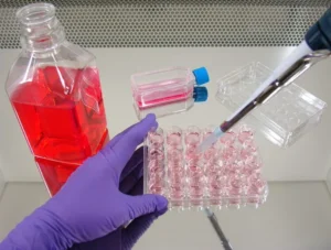
Before testing on the retina, the team needed to ensure that these nanoparticles could safely communicate with living tissue. Using cardiac cells (cardiomyocytes) and cardiac fibroblasts, the researchers demonstrated that focused laser stimulation could induce "calcium-transient release"—a critical step in cellular signaling. By using LED light to pace and synchronize the beating of these cells, the team proved that their material could indeed bridge the gap between light and biological movement.
Phase 3: Retinal Integration and Behavioral Response
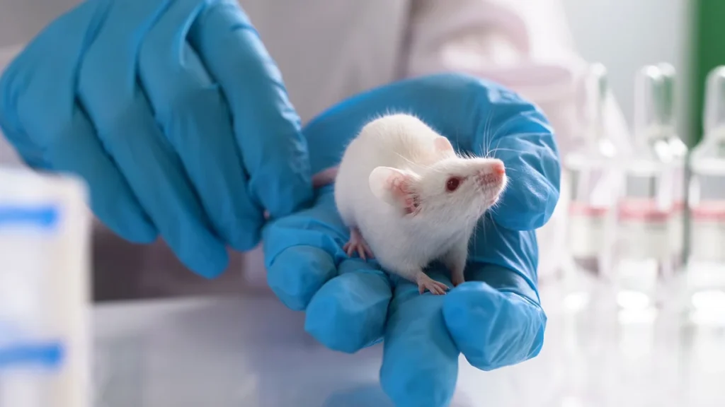
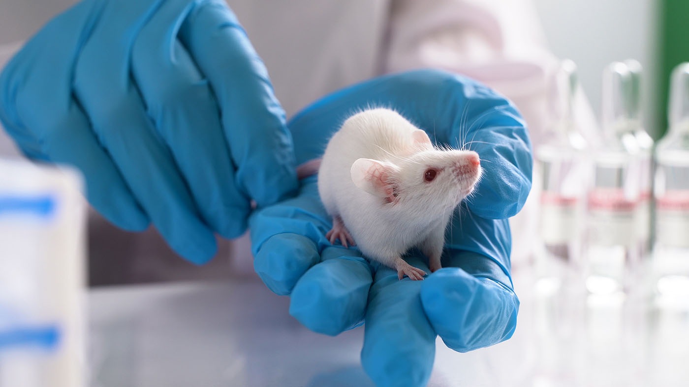
Moving to the eye, the team tested the nanoparticles in mouse models suffering from advanced retinitis pigmentosa. Upon injection, the particles naturally accumulated near the retinal ganglion cells—the vital neurons that relay processed visual information from the retina to the visual cortex of the brain. The results were striking: upon exposure to light, the researchers detected clear activity in the visual cortex, and the mice exhibited behavioral responses to light stimuli, indicating that the signal had successfully traveled from the eye to the brain.
Phase 4: Scaling to Porcine Tissue
To bridge the gap between rodent models and potential human application, the researchers utilized isolated porcine retinal tissue. Under LED photostimulation, the nanoparticles successfully activated retinal ganglion cells, demonstrating that the mechanism remains effective in larger, more complex biological systems.
Supporting Data: Translating Light into Life
The study, titled “Biomimetic graphitic carbon nitride nanoparticles for multiscale photomodulation and therapeutic intervention,” provides a wealth of data regarding the efficiency of these particles.
Key metrics from the study include:
- Targeted Accumulation: The nanoparticles were found to lodge effectively near retinal ganglion cells without the need for complex, targeted delivery vectors.
- Multiscale Signal Propagation: The ability of the particles to trigger calcium signaling across different cell types (cardiac and neural) validates the "universal" nature of the interface.
- Non-Genetic Modification: Unlike traditional optogenetics, which requires the viral delivery of light-sensitive protein genes into the patient’s own cells, this approach avoids permanent genetic alteration, significantly lowering the regulatory and biological risk profile.
- Behavioral Confirmation: The restoration of behavioral responses in blind mice suggests that the visual information being transmitted is not merely noise, but a coherent signal that the brain can recognize as light.
Official Perspectives: The Road Ahead
While the scientific community has hailed the study as a significant milestone, the researchers remain measured in their optimism. The transition from a laboratory bench to a clinical trial is fraught with obstacles.
"Instead of genetically modifying these cells, we use the nanoparticles to create a new connection between light and the nerve cells," Chen noted. "In this way, we are trying to make a blind retina respond to light again."
Collaborators from the University of Chicago, the University of Eastern Finland, and the University of Copenhagen have echoed the sentiment that the next phase of development will focus on the "durability" of the interface. Key questions remaining for the research team include:
- Biocompatibility and Clearance: How long do these particles remain within the vitreous humor of the eye, and are they eventually cleared by the body’s natural metabolic processes?
- Signal Fidelity: Can the "resolution" of the light-to-nerve signal be sharpened? Currently, the response is a general detection of light; for true sight, the brain requires high-fidelity, localized data.
- Long-Term Safety: While initial studies show no adverse reactions, chronic exposure to semiconductor nanoparticles in the delicate environment of the retina requires extensive, long-term toxicological assessment.
Implications for the Future of Ophthalmology
The implications of this research extend far beyond the treatment of retinitis pigmentosa. If this "wireless interface" technology can be perfected, it could provide a blueprint for treating a wide array of neurodegenerative conditions.
A New Class of Retinal Prosthesis
Currently, retinal prostheses are bulky, expensive, and invasive. A liquid-based, injectable solution would represent a "minimally invasive" revolution. Patients could potentially receive a simple injection rather than a high-risk surgical implant, making the therapy accessible to a much broader demographic of patients worldwide.
Beyond Vision
The ability to stimulate nerve cells using light-sensitive materials could eventually be applied to other parts of the nervous system. From treating spinal cord injuries to modulating nerve signals in chronic pain patients, the concept of "light-activated cellular interfaces" opens a new door in bio-electronic medicine.
The Challenge of Resolution
For vision specifically, the hurdle remains the human brain’s requirement for high-resolution input. The current study shows that the retina can "see" light, but the next step is determining if it can "see" shapes, motion, and color. Future iterations of the nanoparticles may need to be coupled with specialized eyewear—smart glasses that process visual data and project it into the eye at specific wavelengths to maximize the stimulation of the nanoparticles.
Conclusion: A Glimmer of Hope
The work of Dr. Menglin Chen and her international cohort represents the best of modern science: a fusion of materials engineering, biology, and clinical ambition. By looking to nature—specifically the mechanism of photosynthesis—the team has managed to solve a problem that has baffled scientists for decades.
While we are not yet at the stage of restoring 20/20 vision to those who have lost it, the study establishes a crucial fact: the architecture of the human eye is more resilient than we once thought. Even in the darkness of degeneration, the biological "wiring" remains. With the right bridge, we may soon find a way to turn the lights back on.
As the research moves into the next phase of preclinical development, the eyes of the medical world will be fixed on Aarhus University. The "solar cell" for the human eye is no longer a science-fiction concept; it is an active, evolving technology that promises to redefine the boundaries of human vision.