Unlocking the Embryonic Blueprint: Scientists Use DNA "Typewriters" to Map Mammalian Life
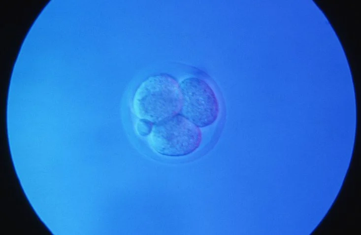
In a landmark achievement for developmental biology, researchers have successfully reconstructed the cellular "family trees" of mammalian embryos, overcoming a hurdle that has long frustrated science: the inability to observe development in real-time within the opaque, inaccessible environment of the uterus. By repurposing the genetic material of living cells as a high-fidelity recording medium, two independent teams have provided the most detailed look yet at how a single fertilized egg transforms into a complex, multi-organ organism.
The breakthrough, detailed in companion studies published in Science and Cell, represents a shift in how we understand the "lineage" of life. By effectively installing a molecular "black box" recorder into the genome of a mouse embryo, scientists have moved from static snapshots of biology to dynamic, chronological maps of cellular decision-making.
The Challenge of the Womb: Why Development Remained a Mystery
For decades, the field of developmental biology has relied on two primary, yet flawed, methodologies. The first is live-cell imaging—the gold standard for transparency, which allows researchers to watch cells divide under a microscope. However, this is largely limited to organisms that are transparent or develop outside of a mother, such as the roundworm C. elegans or the zebrafish. Mammalian development, protected deep within the uterus, is inherently shielded from such observation.
The second methodology, single-cell genomics, offers a high-resolution "snapshot" of what a cell is doing at a precise moment in time. But these techniques are destructive; they require killing the cells to analyze their RNA or protein content. Consequently, researchers could see where a cell ended up, but they could never truly know the path it took to get there.
The new studies, led by researchers at the University of Washington School of Medicine, Dartmouth College, and the Whitehead Institute/MIT, bridge this gap. By embedding "recording systems" into the genome, the researchers ensured that as a cell divided, it left behind a permanent, sequential record of its ancestry that could be read long after the embryo had developed.
Chronology of a Breakthrough: The DNA Typewriter
The Science study, spearheaded by co-senior authors Jay Shendure, PhD, of the University of Washington, and Chengxiang Qiu, PhD, of Dartmouth College, utilized a technology known as the "DNA Typewriter."
Developing the Tape
The concept, originally developed by Shendure and Junhong Choi, PhD, works by modifying a specific segment of the mouse’s DNA to act as a "recording tape." When a fertilized mouse egg is injected with this molecular machinery, the system begins to "type" edits into the genome.
As the zygote divides, these edits are passed down to all daughter cells. Because the system is designed to record sequentially, every subsequent cell inherits the "history" written by its ancestors, while simultaneously adding its own new "characters" to the record.
The Mapping Process
In their experiment, the team attempted to inject 100 fertilized eggs, eventually yielding 10 embryos suitable for high-resolution analysis. By examining the DNA sequences of cells within these embryos, the researchers could look for shared "typos" or edits. If two cells possessed the same initial recording, they were proven to share a common ancestor from the earliest stages of development.
This allowed the researchers to trace almost every cell in the developed embryo back to the very first two cells formed after the initial cleavage. Surprisingly, while one of these two founding cells produced a larger number of descendants, both branches contributed equally to the diversity of cell types in the mature embryo, suggesting a robust and balanced developmental program.
Supporting Data: Mapping the Landscape of Cell Fate
While the Science study focused on the "DNA Typewriter," the Cell study took a complementary approach. Led by Jonathan Weissman, PhD, at the Whitehead Institute, the team utilized a system called "PEtracer," which leverages prime-editing technology to track the lineage of over 1.4 million cells across 16 mouse embryos.
Deciphering Cell Commitment
The combined data from both studies have yielded profound insights into how and when cells "commit" to their identities. Traditionally, it was thought that cell differentiation might be a coordinated, global event. However, these lineage maps reveal that cell identity is not fixed in a single step. Instead, different lineages become restricted on staggered, distinct schedules.
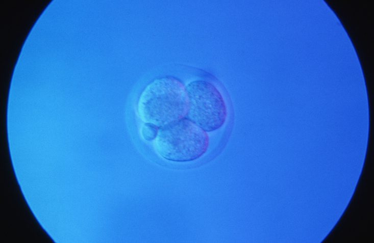
For example, the data showed that blood and retinal cells commit to their roles relatively early in the developmental process. In contrast, cells destined to form the skin’s outer layers remain flexible for a longer period, committing to their final identity much later. This "staggered commitment" provides a new framework for understanding the timing of organogenesis.
Advantages Over Previous Systems
Earlier lineage-tracing techniques often suffered from "molecular fatigue." They relied on DNA-cutting enzymes that were inherently messy; they often caused collateral damage to the genome, erased previous records, or ran out of "space" to record new events. The DNA Typewriter and PEtracer systems solve these issues by:
- Avoiding DNA Severance: The systems write without fully breaking the DNA strand, preserving the integrity of the genome.
- Sequential Precision: Unlike previous methods that left unordered marks, these systems record in a chronological, orderly fashion, allowing researchers to reconstruct the "when" of development with high temporal resolution.
Official Responses and Expert Perspective
The implications of this work are being hailed as a "coming of age" for synthetic biology. Dr. Jay Shendure, an investigator with the Howard Hughes Medical Institute, highlighted the paradigm shift during a recent presentation of the findings.
"Most biological measurements are based on either live imaging, which is limited by the fact that most animal tissues are not transparent, or genomics, which are destructive and only measure a single timepoint," Shendure remarked. "Recording techniques like the ones in these studies enable measurements over time, including in settings that we can’t directly visualize."
Dr. Jonathan Weissman, whose team performed the massive 1.4-million-cell analysis in the Cell publication, framed the achievement as a historical milestone. "It’s really proof of principle that we can do what was done with the roundworm in 1983, but for mammals like you and me," Weissman noted.
The reference to the 1983 C. elegans study is significant; that work was the first to map the entire developmental lineage of an organism. For decades, the complexity of mammalian embryos made such a map seem like a distant, perhaps impossible, dream. The success of the DNA Typewriter suggests that the "lineage map" of a mouse—and eventually, perhaps, a human-relevant model—is now within reach.
Implications: From Congenital Disorders to Stem Cell Engineering
The ability to record the history of cells as they form tissues and organs opens a vast new frontier for clinical and basic research.
Understanding Congenital Malformations
Many congenital disorders—conditions present at birth—arise from developmental errors that occur in the first few weeks of life. Because these errors happen in utero, they are essentially invisible to current diagnostic tools. By using DNA-recording systems in models of disease, researchers may finally be able to pinpoint the exact moment when a developmental pathway diverges, potentially leading to new strategies for intervention.
Advancing Regenerative Medicine
Stem cell engineering relies on our ability to "nudge" undifferentiated cells into becoming specific tissue types, such as heart muscle or neurons. However, the process is often inefficient because we do not fully understand the intermediate "states" a cell passes through during differentiation. By providing a high-resolution map of these natural developmental paths, these lineage-tracing technologies provide a "blueprint" that scientists can follow to improve the efficiency and safety of stem cell therapies.
The Future of Predictive Biology
The authors believe this is only the beginning. As these methods scale to include thousands more embryos under a wider variety of environmental and genetic conditions, the goal is to transition from observational biology to predictive biology. By feeding this massive amount of lineage data into machine learning models, scientists hope to create digital "twins" of development. These models could eventually predict how a specific genetic mutation will alter the trajectory of a developing organ, or how a drug might interfere with the delicate choreography of embryogenesis.
Conclusion
The DNA Typewriter and PEtracer represent more than just clever engineering; they represent a fundamental change in our relationship with the biological record. For the first time, we have the tools to treat the genome not just as a static code, but as a dynamic diary of existence. As these technologies mature, the "black box" of the womb is slowly opening, promising a new era where the mysteries of human development are no longer hidden, but read, recorded, and ultimately understood.