Unveiling the Dark Matter of Cancer Signaling: How Custom Nanotech is Redefining ErbB Receptor Dynamics
In the high-stakes world of oncology, the ErbB family of receptors—EGFR, HER2, and HER3—has long been a primary target for pharmaceutical intervention. Yet, for all our clinical success with inhibitors and monoclonal antibodies, one member of the family has remained stubbornly enigmatic: HER3. Often dismissed as a “weak” kinase with little intrinsic signaling power, HER3 has been treated by the research community as a mere sidekick to its more aggressive counterparts.
Now, a groundbreaking study from the Broad Institute of MIT and Harvard, published in Cell, has fundamentally shifted this paradigm. By leveraging a custom-built, long-duration single-molecule imaging platform, researchers have discovered that wild-type HER3 forms remarkably stable homodimers. Far from being a dormant partner, HER3 appears to function as a regulatory reservoir, with cancer-associated mutations actively dismantling these stable pairings to unleash oncogenic signaling.
This revelation was not merely a triumph of biological insight; it was a victory of instrumentation. By engineering upconverting nanoparticle (UCNP) probes that defy the limitations of conventional fluorescence, the team led by Sam Peng has unlocked a window into the cell that was previously shuttered by the rapid “photobleaching” of standard dyes.
The Technical Breakthrough: Seeing Beyond the Blink
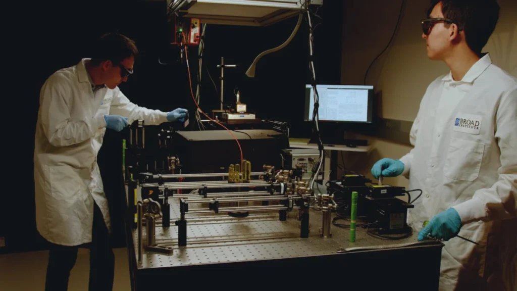
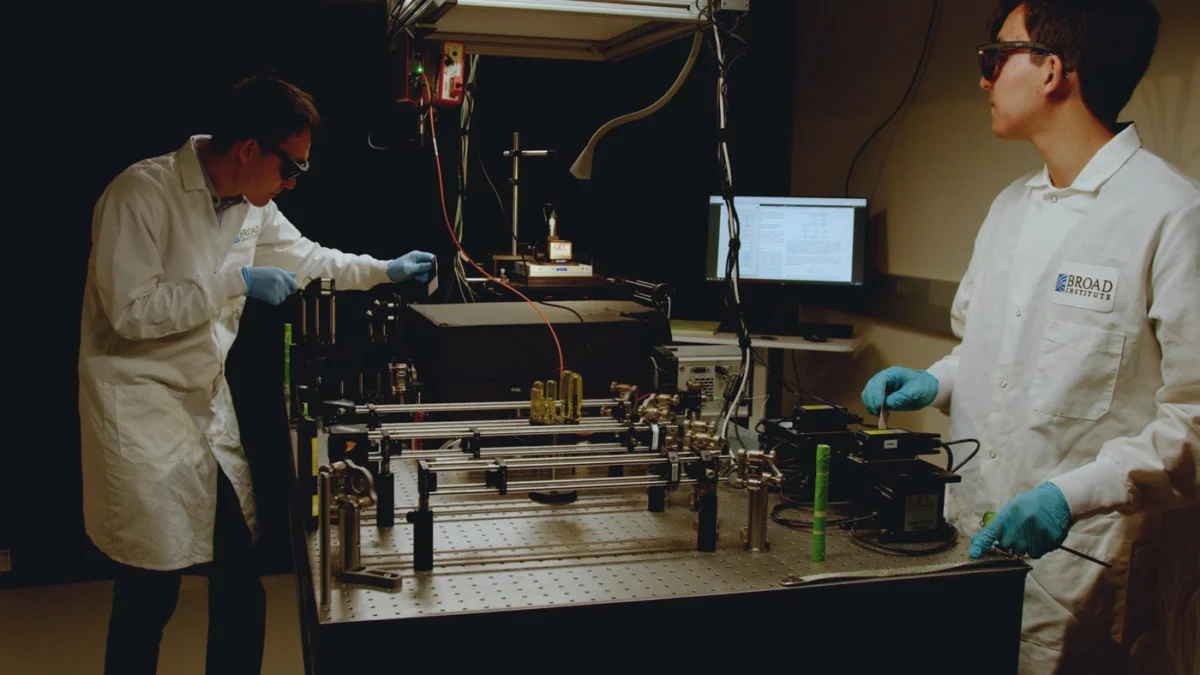
In the corner of Sam Peng’s laboratory, the imaging apparatus looks less like a sleek, off-the-shelf instrument and more like a high-stakes engineering project. Cameras, high-precision lenses, and specialized lasers are bolted directly onto a sprawling metal breadboard. It is an improvised masterpiece—a "Lego-like" architecture of light and detection designed to push the boundaries of spatial and temporal resolution.
The core challenge of live-cell imaging has always been duration. Conventional organic dyes, which act as the “glow-sticks” of the microscopic world, typically burn out within seconds under the intense excitation required to track single molecules.
“If you’re trying to understand a conversation between two people, and I cut that conversation off after two seconds, you are forced to guess the conclusion based on a tiny fragment of interaction,” Peng explains. “That guess is often wrong. By extending our imaging window, we are finally hearing the whole sentence.”
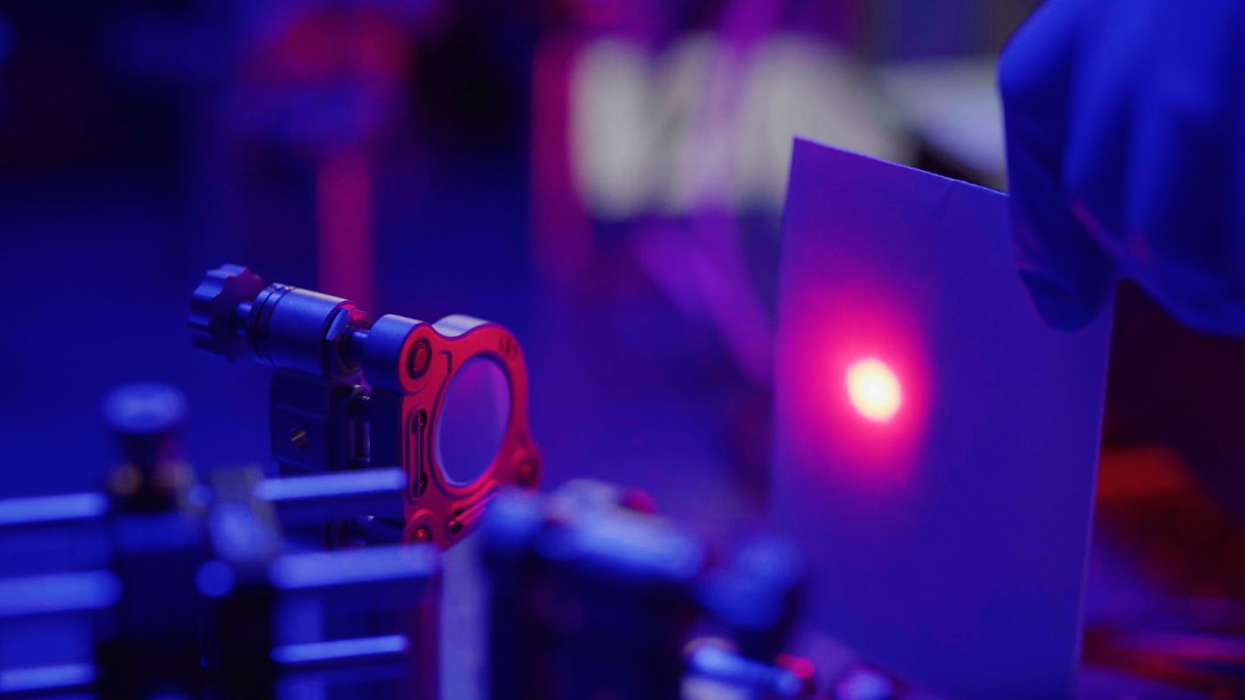
Peng’s team solved this by synthesizing UCNPs—nanoparticles doped with heavy rare earth elements including ytterbium, erbium, and thulium. Unlike conventional fluorophores, these particles do not photobleach. They allow for 100-millisecond resolution tracking of proteins on the surface of live cells for over 16 minutes—a lifetime in the context of molecular signaling. This allows the researchers to witness the dance of EGFR, HER2, and HER3 simultaneously in three distinct colors, providing a panoramic view of receptor behavior that was previously impossible.
Chronology of a Discovery
The path to this discovery was paved with skepticism. Initially, when the team observed HER3 homodimers, they were convinced the data was skewed.
- Initial Hypothesis (2024–2025): The prevailing dogma suggested that HER3, lacking a potent kinase domain, was incapable of autonomous signaling. It was expected to act as a negative control—a protein that should not form homodimers.
- The Validation Phase (Early 2026): Upon observing the “unexpected” pairing, the team repeatedly reran the experiments using diverse labeling strategies to rule out experimental artifacts. The result remained consistent: wild-type HER3 was consistently found in stable homodimer configurations.
- The Mechanistic Breakthrough (Mid-2026): Once the stability was confirmed, the team turned their attention to cancer-associated mutations. They discovered a clear, inverse correlation: whereas mutations in EGFR stabilize its dimers to drive aggressive signaling, mutations in HER3 have the opposite effect. By destabilizing the HER3 homodimer, these mutations effectively “liberate” the receptor, allowing it to seek out and form signaling-active heterodimers with other ErbB members.
- Publication (October 2026): The findings were formally published in Cell under the title, ErbB family receptor dimerization dynamics and dysregulation via long-term single-molecule imaging, marking a new chapter in receptor tyrosine kinase biology.
Supporting Data: The Signaling Landscape
The quantitative data provided by the Peng lab’s imaging rig paints a nuanced portrait of cancer progression. The researchers categorized the receptor behaviors into three distinct patterns:
- EGFR (The Driver): Clinical aggressiveness in EGFR-mutant cancers correlates directly with the stability of the homodimer. The more stable the dimer, the more potent the downstream signaling.
- HER3 (The Reservoir): Wild-type HER3 exists in a sequestered, inactive pool. Oncogenic mutations act as a "key" that releases HER3 from this pool, fueling tumor growth through uncontrolled crosstalk with other receptors.
- HER2 (The Amplified): HER2 mutations only marginally enhance homodimer stability. This aligns with clinical observations that HER2-driven cancers are typically sustained by gene amplification—the sheer volume of receptors—rather than the radical structural shifts seen in EGFR or HER3.
These findings suggest that therapeutic strategies for HER3-driven cancers should perhaps focus on stabilizing the homodimer rather than just inhibiting the monomer, effectively "locking" the receptor in its inactive state.
The Geopolitical Dimension: The Rare Earth Bottleneck
The scientific brilliance of this platform is tethered to the harsh realities of global trade. The UCNP probes rely on heavy rare earth elements (HREEs) like ytterbium and erbium. While these elements are not physically scarce in the Earth’s crust, their refining process is incredibly specialized, and the supply chain is famously concentrated.
China currently controls approximately 85% of global rare earth oxide production, including nearly the entirety of the dysprosium and terbium supply. In 2025, the introduction of export controls on these materials sent shockwaves through industries ranging from EV manufacturing to defense. While the lab’s bench-scale consumption—measured in mere milligrams—is shielded from the immediate price volatility that impacts industrial manufacturers, the research community is watching the U.S.-China trade truce with bated breath.
"We buy our salts in vials, much like a standard lab buys antibodies," Peng notes. "The pink erbium salt is beautiful, but it represents a highly specialized node in the global supply chain."
The current trade truce, extended through early 2027, provides a temporary reprieve. However, the reliance on these materials highlights a growing vulnerability in advanced scientific instrumentation. As these tools become standard for drug discovery, the "expertise gap" may become more significant than the supply gap.
Implications: A New Frontier in Precision Medicine
The implications of the Peng lab’s work extend far beyond the bench. By identifying the regulatory role of the HER3 homodimer, researchers now have a novel target for drug development. Current cancer therapies often struggle with "bypass signaling," where cancer cells switch to alternative pathways when one receptor is blocked. By understanding how HER3 is released from its inactive state, scientists can develop dual-action therapies that prevent this switch.
Furthermore, the success of the custom-built imaging platform serves as a manifesto for the next generation of academic science. It demonstrates that the most profound insights often come not from simply applying existing, commercialized technology, but from the willingness to build the tools required to see what others cannot.
The "Integration Challenge"
Sam Peng emphasizes that the real barrier to progress is not the lack of hardware, but the lack of integrated expertise. To perform this work, a lab must be fluent in four distinct languages:
- Nanomaterial Chemistry: To synthesize the UCNPs.
- Molecular Labeling: To ensure probes bind without disrupting cellular biology.
- Advanced Optics: To build the custom rigs capable of long-duration, high-resolution tracking.
- Computational Biology: To extract meaningful patterns from terabytes of high-dimensional imaging data.
“It’s the integration of the entire pipeline that makes it challenging,” Peng concludes. “You cannot just hire a specialist for one part; you need a team that understands how the optics influence the chemistry, and how the chemistry influences the interpretation of the biological data.”
As the Broad Institute continues to refine these techniques, the hope is that this “long-conversation” approach to single-molecule imaging will reveal the hidden mechanics of other elusive proteins. The era of blink-and-you-miss-it biology is coming to an end, and in its place, a more stable, detailed, and actionable understanding of cellular life is beginning to emerge.
The conversation between these receptors is finally being recorded in full, and for patients waiting for the next generation of targeted therapies, that is the most important development of all.