Capturing the Molecular Movie: Scientists Unveil the Real-Time Synthesis of Penicillin
For over eight decades, the medical world has relied on the potent, life-saving architecture of penicillin—a discovery that fundamentally altered the course of human history. Yet, despite its ubiquity in modern medicine, the intricate, high-speed chemical choreography that allows nature to construct this antibiotic remained largely shrouded in mystery.
Now, in a landmark study published in Nature Catalysis, an international research team led by the University of Oxford has successfully produced a "molecular movie" of penicillin biosynthesis. By utilizing cutting-edge X-ray free-electron laser (XFEL) technology, researchers have captured the fleeting, millisecond-long intermediates of the enzyme isopenicillin N synthase (IPNS). This breakthrough not only resolves a decades-old scientific debate regarding how bacteria assemble the penicillin scaffold but also provides a vital blueprint for the next generation of antibiotic development in an era of mounting antimicrobial resistance.
The Architectural Marvel of the $beta$-Lactam Ring
The potency of penicillin and its derivatives lies in the $beta$-lactam ring—a highly strained, four-membered chemical structure that acts as a molecular "skeleton key." This ring interferes with the synthesis of bacterial cell walls, causing them to rupture and leading to the death of the invading pathogen.
Since the pioneering work of Nobel laureate Dorothy Hodgkin, who solved the structure of penicillin using X-ray crystallography in 1945, scientists have sought to understand the enzymatic machinery behind its creation. IPNS is the biological engine that drives this process; it is an iron-dependent oxygenase that transforms a simple, linear peptide substrate into the fused four-membered $beta$-lactam and five-membered thiazolidine ring system.
However, because these reactions occur in the blink of an eye, traditional structural biology—which relies on static "snapshots" of enzymes—has been unable to capture the intermediate states. The transition from a linear molecule to a complex, cyclic antibiotic involves delicate shifts in shape and high-energy chemical states that vanish almost as quickly as they appear.
Chronology of a Breakthrough: From Static to Dynamic Biology
The road to this "molecular movie" involved a massive interdisciplinary effort, bringing together structural biology, quantum chemistry, spectroscopy, and advanced engineering. The collaboration included researchers from the University of Oxford, Diamond Light Source, Lawrence Berkeley National Laboratory, PAL-XFEL, and the SLAC National Accelerator Laboratory.
The Experimental Setup
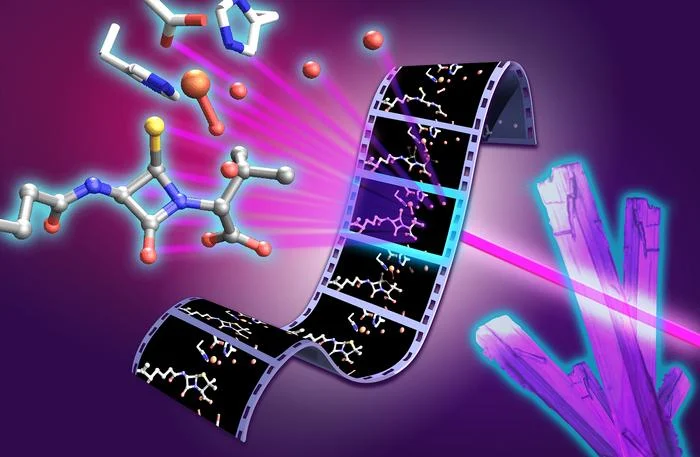
To overcome the limitations of static imaging, the team developed an ingenious experimental pipeline. The process began with the creation of thousands of microcrystals containing the IPNS enzyme. These crystals were deposited onto a moving, two-millimeter-wide tape. As the tape advanced into an oxygen-rich chamber, oxygen diffused into the crystals, triggering the catalytic reaction across the entire sample simultaneously.
By carefully modulating the speed of the tape, the researchers created a "time-resolved" sequence. They could determine exactly how long the reaction had been running before the crystal reached the interaction point, where an ultrafast XFEL pulse captured an atomic-resolution snapshot. By stitching together thousands of these individual frames, the researchers created a continuous, frame-by-frame film strip of the chemical reaction as it unfolded in real time.
Capturing the Fleeting Stages
The movie reveals, for the first time, the transition states that occur during the conversion of the peptide substrate into penicillin. Key highlights from the footage include:
- The Thioaldehyde Intermediate: A transient state formed just milliseconds before the $beta$-lactam ring begins to close.
- The Monocyclic $beta$-Lactam Intermediate: The first completed ring structure on the path to the final scaffold.
- Ferryl Iron Species: The study provides visual evidence of the high-valent iron species at the heart of the enzyme’s active site, demonstrating how the iron atom coordinates the electron transfer necessary for ring formation.
These snapshots revealed that the enzyme is not a passive scaffold; rather, it performs subtle, rhythmic movements that guide the chemistry, acting as a molecular conductor that ensures the reaction proceeds with near-perfect precision.
Supporting Data: The Role of Water and Enzyme Motion
One of the most surprising findings to emerge from the XFEL data is the critical, previously underestimated role of water molecules residing within the enzyme’s active site. For forty years, the mechanism of IPNS was debated, with many models failing to account for the exact sequence of proton transfers.
The study confirms that these internal water molecules are not merely structural "filler." Instead, they are active participants in the reaction, facilitating the proton transfers required to stabilize the intermediate states. The researchers noted that the enzyme’s ability to "sculpt" the environment—positioning water molecules to donate and accept protons at the precise moment—is what allows such a complex chemical transformation to occur in a single, efficient step.
This discovery provides the first empirical resolution to a long-standing mechanistic debate. It demonstrates that the enzyme’s structural flexibility is perfectly synchronized with the electronic requirements of the iron catalyst, confirming that enzyme shape and chemistry function as a unified, inseparable system.
Official Responses: A New Era for Antibiotic Design
The implications of this study are profound, particularly given the escalating global crisis of antimicrobial resistance (AMR). According to the World Health Organization, one in six bacterial infections is now resistant to common antibiotics, and the pipeline for new, effective drugs has slowed to a trickle.
"Penicillin has shaped modern medicine, but there is still much to learn about how nature builds this important antibiotic structure," said Professor Christopher Schofield of the University of Oxford, co-senior author of the study. "By capturing these fleeting steps, we can better understand how enzymes control complex chemistry with remarkable precision. As rates of antimicrobial resistance continue to rise, understanding this process will ultimately help us make existing antibiotics more efficient and design new antibiotic structures."
Dr. Allen M. Orville, group leader of the XFEL Hub at Diamond Light Source, emphasized the collaborative nature of the success. "The results show the power of combining complementary expertise and X-ray techniques to move beyond static structures and reveal how enzymes work in real time. It is a great example of how time-resolved structural biology can uncover new principles of enzyme function and, ultimately, inform the design of new catalysts and therapeutics."
Dr. Patrick Rabe, the study’s first author and a Wellcome Career Development Award Investigator, highlighted the practical application of the findings: "The ability to capture structural snapshots over milliseconds to seconds allows us to connect enzyme motion, iron chemistry, and water-mediated proton transfer. This gives us a much richer view of how IPNS controls a difficult chemical transformation. By understanding this process in atomic detail, we can begin to think about engineering these enzymes to produce new or improved antibiotic scaffolds."
Implications for Future Therapeutics and Beyond
The reach of this research extends well beyond the production of penicillin. IPNS belongs to a massive family of iron-dependent oxygenase enzymes that play crucial roles in human biology, including oxygen sensing and metabolic regulation. By mastering the ability to observe these enzymes in action, scientists have opened a new door for enzyme engineering.
Revolutionizing Drug Discovery
If scientists can understand how nature builds a $beta$-lactam ring with such efficiency, they can theoretically replicate or modify this process in a laboratory setting. This opens the door to:
- Custom-Designed Antibiotics: Modifying IPNS to produce novel, "custom" scaffolds that can bypass the resistance mechanisms evolved by modern bacteria.
- Bio-Catalysis: Utilizing modified enzymes to manufacture complex pharmaceuticals in a more sustainable, energy-efficient way, reducing the need for harsh synthetic solvents.
- Targeting Human Disease: Many enzymes in the human body function through similar mechanisms. Insights into how IPNS controls its iron-based active site could inform the development of drugs that target enzymes involved in cancer, neurodegeneration, and inflammatory diseases.
The Power of Time-Resolved Structural Biology
The success of this experiment marks a transition in the biological sciences. For decades, structural biology was akin to examining a fossil—a static record of what once was. Time-resolved XFEL methods turn that fossil into a live performance. This shift allows researchers to observe the transition states that are the "heart" of biological function.
As the research team looks toward the future, they anticipate that this methodological framework—combining XFEL snapshots with computational modeling—will become a standard approach for analyzing enzyme catalysis. By revealing the "how" behind the "what," scientists are finally gaining the ability to manipulate the machinery of life with the same precision that evolution has used for millions of years.
In conclusion, the "molecular movie" of penicillin biosynthesis is more than a technical triumph; it is a vital step toward securing the future of medicine. By learning the language of the enzyme, researchers are better equipped to combat the silent, growing threat of antibiotic resistance, potentially ushering in a new generation of therapeutics designed with the same elegance and efficiency as the very antibiotics that first saved millions of lives in the 20th century.